Hi-Affi™ In Vitro Cell based Glycoprotein Hormone Receptor Functional Assay Service
The Importance of Glycoprotein Hormone Receptors
Glycoprotein hormone receptors (GPHRs) are rhodopsin-like GPCRs with a substantial extracellular N-terminal region that is essential for hormone recognition and binding. TSH receptor (Thyrotropin), FSH receptor (Follitropin), and LH receptor (Lutropin) are three types of GPHRs. Although their major physiological function is to support gonadal progress, several studies have revealed that mutations in these receptors cause the emergence of certain disorders that are difficult to treat. Whereupon, there is an urgent need for an in-depth interpretation of GPHRs.
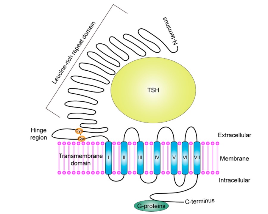 Fig.1 The schematic diagram of the folded protein structure of TSHR.1
Our Hi-Affi™ In Vitro Cell-Based Glycoprotein Hormone Receptor Functional Assay Service
To boost the development of drug discovery, Creative Biolabs provides a simple and effective Hi-Affi™ in vitro cell-based glycoprotein hormone receptor functional assay service for global customers.
In our Hi-Affi™ in vitro cell-based glycoprotein hormone receptor functional assay service, we integrate in vitro cell-based assay with high sensitivity and effectiveness to glycoprotein hormone receptors functional assay. Through investigating changes in second messenger concentrations and downstream calcium fluxes resulting from receptor activation, this assay allows us to identify compounds and their ability to modify glycoprotein hormone receptors. Together with our high-quality comprehensive customized services, Creative Biolabs' Hi-Affi™ in vitro cell-based glycoprotein hormone receptor functional assay service can satisfy the diverse demands of global customers.
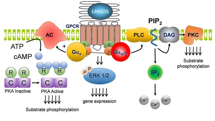 Fig.2 Signaling pathways downstream of LHCGR.2
Benefit for You
Our Hi-Affi™ in vitro cell-based glycoprotein hormone receptor functional assay service can benefit you with:
 Fig.3 Benefit of our Hi-Affi™ in vitro cell-based glycoprotein hormone receptor functional assay service.
Case Study
Representative Data 1: Key signaling pathways in HEK-293 cells recombinantly expressing LHCGR (HEK-LHCGR) were examined to confirm gonadotropin bioactivity. Cells were challenged with either hCG or LH and compared to wild-type HEK-293 cells (HEK-WT) in parallel studies.
- cAMP assay: Intracellular cAMP levels increased significantly in HEK-LHCGR cells after 5 minutes of exposure to either hCG or LH. Inductions were concentration-dependent, with pEC50 values for hCG of 9.671 (213 pM) and LH of 9.370 (426 pM), with peak responses at 10 nM. HEK-WT cells, on the other hand, did not react.
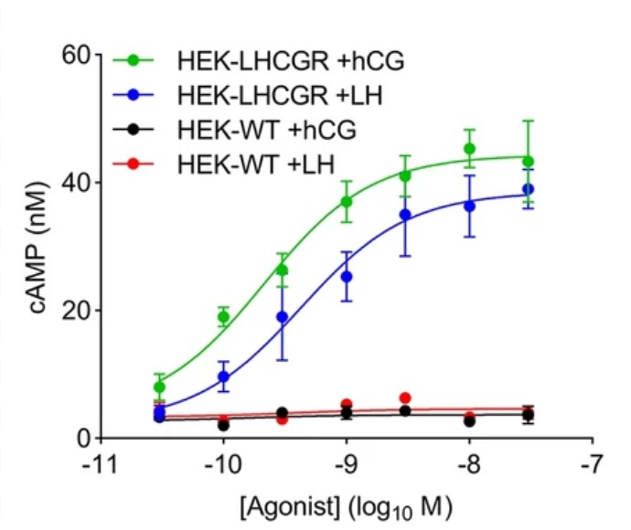 Fig.4 cAMP responses to hCG and LH in HEK-WT and HEK-LHCGR cells.2
- Ca2+ assay:, a Ca2+-sensitive dye, was also added to HEK cells before they were challenged with high doses of hCG or LH (1 M). In HEK-LHCGR cells, both agonists induced strong Ca2+ responses, but not in HEK-WT cells.
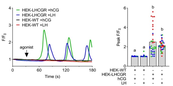 Fig.5 Ca2+ signals in HEK-WT and -LHCGR cells after hCG and LH treatment, respectively.2
- ERK phosphorylation assay: Both ligands resulted in a significant and prolonged elevation of phosphorylated (active)-ERK in HEK-LHCGR cells, but HEK-WT cells did not react.
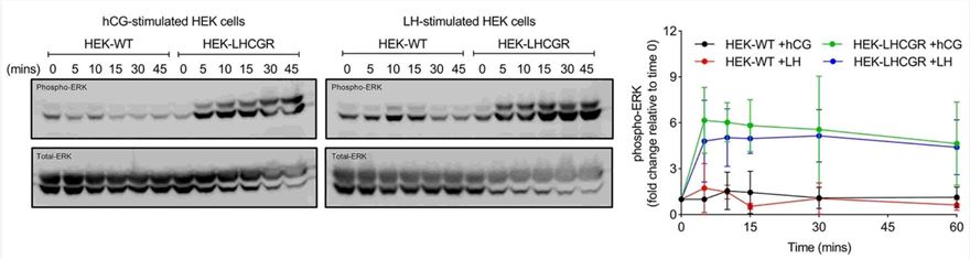 Fig.6 The effects of hCG and LH on phospho-ERK levels in wild-type and LHCGR-expressing HEK cells.2
As an industry leader in GPCR functional assays, Creative Biolabs is dedicated to providing every customer with the most advanced techniques as well as top-ranking services. For more details about our Hi-Affi™ in vitro cell-based glycoprotein hormone receptor functional assay service, please don't hesitate to get in touch with us. All of us here are eager to get to work with you right away.
References
-
Chu, Yu-De, and Chau-Ting Yeh. "The molecular function and clinical role of thyroid stimulating hormone receptor in cancer cells." Cells 9.7 (2020): 1730.
Distributed under Open Access License CC BY 4.0. The original image was modified by extracting and using part B, and the title was changed to "The schematic diagram of the folded protein structure of TSHR". -
Mann, O. N., et al. "Expression and function of the luteinizing hormone choriogonadotropin receptor in human endometrial stromal cells." Scientific Reports 12.1 (2022): 8624.
Distributed under Open Access License CC BY 4.0. The original image Figure 2 was modified by extracting and using parts a, b, c, and d, and their titles were changed to "Signaling pathways downstream of LHCGR", "cAMP responses to hCG and LH in HEK-WT and HEK-LHCGR cells", "Ca2+ signals in HEK-WT and -LHCGR cells after hCG and LH treatment, respectively", and "The effects of hCG and LH on phospho-ERK levels in wild-type and LHCGR-expressing HEK cells", respectively.
For Research Use Only.