

The exosome controlled apoptosis regulation assay service at Creative Biolabs is designed to support in vitro research focused on how exosomes influence programmed cell death. Apoptosis is a tightly controlled biological process that plays a central role in maintaining cellular homeostasis. Alterations in apoptotic responses often reflect changes in intracellular signaling pathways triggered by external stimuli, including exosome mediated communication.
Creative Biolabs provides carefully designed assays to evaluate both early and late stages of apoptosis following exosome exposure. By combining sensitive detection methods with controlled experimental conditions, this service enables clear differentiation between survival promoting and apoptosis inducing effects. Experimental parameters are tailored based on cell type, treatment duration, and research objectives to ensure meaningful and reproducible outcomes.
| Step | Detail |
|---|---|
| Step 1. Experimental Design and Objective Clarification | Creative Biolabs begins by defining whether the study aims to assess apoptosis induction, inhibition, or modulation under specific conditions. Cell type, treatment duration, and expected response patterns are carefully considered. |
| Step 2. Cell Preparation and Culture Optimization | Recipient cells are cultured under standardized conditions to ensure stable baseline apoptosis levels. Cell health and density are controlled to reduce variability prior to treatment. |
| Step 3. Exosome Treatment and Condition Setup | Exosomes are applied at defined concentrations. If required, apoptosis inducing stimuli such as chemical stressors can be introduced to evaluate protective or regulatory effects. |
| Step 4. Apoptosis Detection and Staining | Cells are processed using validated staining methods targeting key apoptotic markers. These may include membrane changes, DNA fragmentation, or caspase activation depending on study design. |
| Step 5. Data Acquisition and Quantification | Flow cytometry or imaging based systems are used to quantify apoptotic cell populations. Early and late apoptotic stages are distinguished to provide detailed insight into response dynamics. |
| Step 6. Data Analysis and Interpretation | Creative Biolabs performs normalization against control groups and provides statistical analysis. Results are organized to highlight differences in apoptotic response between experimental conditions. |
| Step 7. Reporting and Data Delivery | Final deliverables include raw data, processed outputs, and clear documentation of methods. Data presentation supports integration with other functional assays. |
To refine any step of the workflow or include additional markers, clients are welcome to reach out to the Creative Biolabs technical support team.
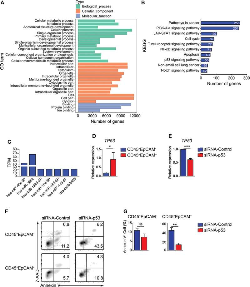 Fig.1 HCC827 cell-derived exosomes promote CD45+EpCAM+ cell apoptosis via the p53 pathway.1
Apoptosis analysis reveals whether exosomes promote survival or contribute to programmed cell death pathways.
Under induced conditions, this assay helps determine how exosomes influence cellular responses to environmental or chemical stress.
Changes in apoptotic markers provide insight into signaling pathways that govern cell fate outcomes.
Quantitative apoptosis data strengthen conclusions in studies focused on intracellular regulatory networks.
Researchers seeking to incorporate apoptosis analysis into broader experimental designs are encouraged to contact Creative Biolabs for tailored advice.
 Detection of Multiple Apoptosis Stages
Assays distinguish early and late apoptotic events, providing a more complete understanding of cellular response.
Flexible Experimental Conditions
Both basal and induced apoptosis models can be implemented depending on research goals.
Quantitative and Qualitative Readouts
Data include numerical measurements as well as visual confirmation when imaging approaches are applied.
Integration with Mechanistic Studies
Apoptosis data can be correlated with viability, proliferation, or signaling assays to strengthen biological interpretation.
For recommendations on assay configuration or marker selection, Creative Biolabs specialists are available for consultation.
For researchers interested in similar assay outcomes, Creative Biolabs welcomes direct inquiries to discuss project needs.
Careful evaluation of apoptosis provides critical insight into how exosomes influence cell fate decisions. Through well controlled experimental design and reliable detection methods, Creative Biolabs delivers data that support detailed mechanistic research. For investigators seeking a dependable partner in apoptosis analysis, our team is ready to assist with customized study design and ongoing technical support.
A: Creative Biolabs uses marker based detection methods that identify membrane changes and DNA integrity differences. These allow separation of early stage apoptotic cells from those in later stages. Data analysis is structured to clearly present these distinctions.
A: Yes. By introducing controlled stressors, the assay can evaluate whether exosome treatment reduces apoptosis levels compared to untreated controls. This approach is commonly used to study cytoprotective mechanisms.
A: Sensitivity depends on cell type, baseline apoptosis levels, and the selected detection method. Creative Biolabs optimizes staining protocols and experimental timing to ensure reliable detection of changes.
A: In many cases, apoptosis assays can be aligned with viability or proliferation studies. Creative Biolabs can advise on experimental design to ensure compatibility and meaningful data integration.
A: Control groups typically include untreated cells and, when relevant, cells exposed to apoptosis inducing conditions without exosome treatment. These controls provide reference points for interpreting experimental effects.
Reference