
Cell adhesion governs how cells attach to substrates and interact with neighboring cells, shaping tissue organization and signaling responses. Exosomes can influence adhesion by transferring proteins, lipids, and nucleic acids that modulate surface receptors and extracellular matrix interactions. The exosome promoted cell adhesion assay service from Creative Biolabs is designed for in vitro research to quantify how exosome exposure alters adhesion strength, attachment kinetics, and cell spreading behavior.
Creative Biolabs establishes assay conditions that reflect the biological context of each study, taking into account cell type, matrix composition, and expected adhesion dynamics. By combining quantitative readouts with controlled experimental setups, this service enables researchers to connect exosome mediated signaling with measurable changes in cellular attachment.
| Step | Detail |
|---|---|
| Step 1. Study Design and Adhesion Model Selection | Creative Biolabs evaluates the research objective to determine appropriate substrates such as collagen, fibronectin, or synthetic coatings, along with suitable cell models. |
| Step 2. Surface Preparation and Plate Conditioning | Culture surfaces are coated and stabilized under standardized conditions to ensure consistent binding environments across replicates. |
| Step 3. Cell Seeding and Baseline Attachment Control | Cells are seeded at defined densities and allowed to reach a controlled baseline attachment state prior to exosome exposure. |
| Step 4. Exosome Treatment Strategy | Exosomes are introduced either before seeding or after initial attachment depending on whether early adhesion or reinforcement effects are being studied. |
| Step 5. Adhesion Measurement and Washing Steps | Non-adherent cells are removed through gentle washing, followed by quantification of attached cells using staining, imaging, or colorimetric methods. |
| Step 6. Imaging and Morphological Assessment | Cell spreading, shape changes, and focal adhesion patterns are recorded to complement quantitative adhesion data. |
| Step 7. Data Analysis and Reporting | Creative Biolabs provides normalized adhesion metrics, statistical comparisons, and visual outputs for clear interpretation. |
To adjust workflow details or include additional matrix conditions, you are welcome to contact Creative Biolabs scientists directly.
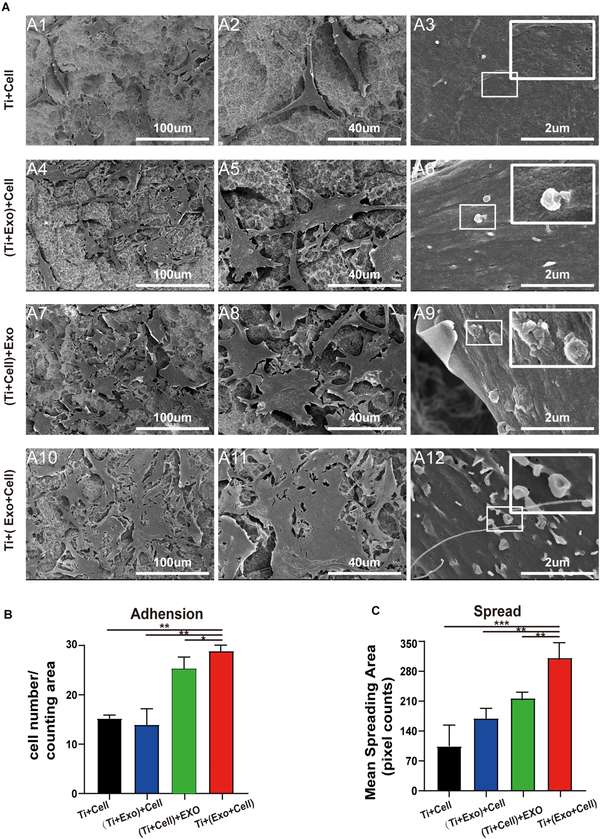 Fig.1 SEM images of rBMSCs with various exosome treatments.1
Adhesion assays reveal how exosomes alter the ability of cells to interact with their surrounding environment.
Changes in adhesion often reflect regulation of integrins and related signaling pathways.
Different exosome preparations can be evaluated for their relative impact on adhesion behavior.
Adhesion data provide functional evidence for intercellular signaling mediated by exosomes.
Researchers planning adhesion related experiments can reach out to Creative Biolabs for detailed input.
 Multiple Adhesion Substrate Options
Supports a range of extracellular matrix coatings to reflect different biological environments.
Quantitative and Morphological Readouts
Combines numerical adhesion data with imaging based confirmation of cell behavior.
Flexible Treatment Timing
Allows investigation of both initial attachment and post-attachment strengthening effects.
Adaptation to Diverse Cell Types
Protocols are optimized for adherent, semi-adherent, and specialized cell models.
For recommendations on assay configuration, Creative Biolabs experts are ready to assist.
For similar project experiences, connecting with Creative Biolabs is highly encouraged.
Understanding how exosomes influence cell adhesion provides a valuable link between molecular signaling and functional cellular behavior. With carefully controlled conditions and reliable quantification strategies, Creative Biolabs delivers data that support detailed in vitro research. For customized assay design or to initiate your project, reaching out to Creative Biolabs is the best next step.
A: The selection depends on the biological relevance to your study. Creative Biolabs reviews the cell type and research objective to recommend coatings that best reflect physiological or experimental conditions.
A: Yes. Quantitative adhesion measurements are complemented by imaging analysis, allowing differentiation between initial attachment and subsequent spreading behavior.
A: Time course designs can be incorporated to monitor how adhesion evolves after exosome exposure, providing insight into early versus sustained effects.
A: Standard controls include untreated cells and, when relevant, matrix only conditions. These controls help establish baseline adhesion levels for comparison.
A: Yes. Adhesion results can be integrated with migration, proliferation, or signaling studies to strengthen mechanistic conclusions.
A: Cell density can influence attachment behavior. Creative Biolabs carefully optimizes seeding conditions to ensure consistent and interpretable results.
Reference