
Wound healing is a complex process that involves cell migration, proliferation, and extracellular matrix remodeling. Exosomes play an important role in intercellular communication during this process, and understanding their impact can shed light on their therapeutic potential in regenerative medicine. Creative Biolabs' assay is designed to quantify how exosome treatment affects the collective movement and closure of a wound area in cell monolayers. This service provides valuable insights into the dynamics of cellular responses to injury, highlighting the role of exosomes in enhancing or modulating tissue repair mechanisms.
| Step | Detail |
|---|---|
| Step 1. Study Objective Definition and Experimental Design | Creative Biolabs collaborates with researchers to define the goal of the study, whether it's evaluating exosome-driven enhancement of wound healing or investigating the underlying molecular pathways involved. Cell types and experimental conditions are selected based on the specific needs of the research. |
| Step 2. Cell Culture and Pre-Conditioning | Cell monolayers are cultured to confluence and prepared for the wound healing assay. If required, cells may be pre-treated with exosomes before the wound is introduced. This step ensures that any changes in healing are directly related to exosome exposure. |
| Step 3. Wound Creation and Exosome Treatment | A defined wound is created using a scratch method, and exosomes are applied at predetermined concentrations. Treatment duration and timing are adjusted to match the research objectives, allowing for real-time tracking of the wound closure process. |
| Step 4. Monitoring and Imaging | The wound closure is monitored using time-lapse imaging or other microscopy techniques to track cell migration into the wound area. Images are captured at multiple time points to assess the rate and extent of wound closure. |
| Step 5. Data Analysis and Quantification | The rate of wound closure is calculated based on the percentage of the wound area covered by cells over time. Creative Biolabs also evaluates the consistency of healing between replicates, ensuring the reliability of the data. |
| Step 6. Reporting and Data Delivery | Creative Biolabs delivers processed data, including graphical representations of wound closure kinetics, statistical analysis, and images. These results provide clear insights into the effects of exosomes on wound healing processes. |
For any adjustments or additional analysis options, Creative Biolabs' technical support team is ready to assist with further customization.
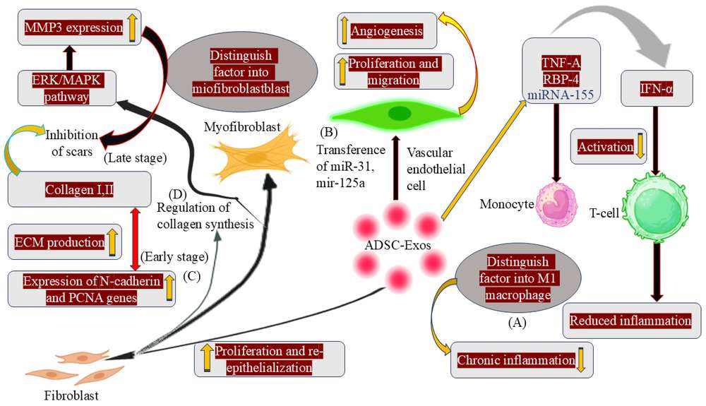 Fig.1 ADSC-Exosomes in Wound Healing and Inflammation Regulation.1
This assay helps investigate how exosomes influence cell migration and collective behavior in response to injury. It is particularly valuable for studying the healing process in tissue repair and regeneration.
Exosome treatments can be evaluated for their potential to enhance wound healing, providing insight into their regenerative properties. This has implications for tissue engineering and wound care.
Understanding how exosomes enhance wound healing can reveal the molecular signaling pathways involved in tissue repair and regeneration, supporting broader studies on intercellular communication.
This assay provides a model for studying how cells respond to mechanical injury and how exosomes modulate cellular reactions to facilitate tissue repair.
For help with specific assay configurations or to discuss the potential applications of this assay for your study, Creative Biolabs' scientific team is available to offer guidance.
 Real-Time Monitoring
Wound healing is tracked in real-time, allowing researchers to observe dynamic changes in cell movement and tissue repair over time.
Flexible Experimental Design
Protocols can be customized to include different exosome sources, cell types, or treatment durations to align with the specific objectives of the study.
Quantitative and Visual Analysis
Both quantitative data (wound closure percentage) and visual evidence (time-lapse images) are provided to support comprehensive interpretation.
Adaptation to Different Cell Types
This service supports a variety of cell models, including epithelial, fibroblasts, and other relevant cell types, based on the specific research focus.
For more information on customizing your study or adding other functional assays, Creative Biolabs' expert team is available for consultation.
For similar project outcomes, Creative Biolabs invites direct communication to discuss your experimental needs.
Understanding the role of exosomes in wound healing is crucial for advancing research in tissue repair and regeneration. Creative Biolabs provides a reliable and reproducible assay to evaluate exosome-driven effects on wound closure, offering valuable data for researchers. For customized solutions or further assistance, please feel free to reach out to our technical support team.
A: The assay generally takes 24-48 hours, depending on the time required for cells to migrate into the wound area and close it. Time-lapse imaging allows for monitoring the progress at multiple intervals.
A: Yes. This assay can be used to measure both the enhancement and inhibition of wound healing. Researchers can compare the effects of different exosome preparations or inhibitors to assess their impact on tissue repair.
A: The wound closure is measured by calculating the area of the wound that has been covered by migrating cells at each time point, relative to the original wound area.
A: The assay can be performed using various cell types, including epithelial cells, fibroblasts, and other relevant models for wound healing. The cell type is selected based on the research question and experimental design.
Reference