
The exosome labeling and receptor cell uptake assay service at Creative Biolabs is designed to support mechanistic in vitro research focused on intercellular communication. This service enables researchers to visualize, quantify, and interpret how exosomes interact with recipient cells under controlled laboratory conditions. By combining optimized labeling strategies with robust uptake detection systems, Creative Biolabs helps generate reproducible data that clarifies binding efficiency, internalization dynamics, and intracellular distribution patterns.
Accurate assessment of exosome uptake is essential for understanding functional outcomes observed in downstream assays. Differences in receptor expression, membrane composition, and experimental timing can significantly influence internalization behavior. Creative Biolabs applies carefully validated protocols tailored to the biological source of exosomes and the characteristics of recipient cells.
| Step | Detail |
|---|---|
| Step 1. Experimental Design Consultation | Creative Biolabs begins with a detailed review of the exosome source, isolation method, recipient cell type, and research objectives. Parameters such as labeling approach, incubation conditions, and detection method are defined to align with the study goals. |
| Step 2. Exosome Labeling Optimization | Exosomes are labeled using validated fluorescent dyes or reporter based systems selected according to experimental needs. Labeling efficiency, background signal, and vesicle integrity are assessed to ensure that fluorescence reflects true vesicle behavior rather than artifacts. |
| Step 3. Recipient Cell Preparation | Target cells are cultured under standardized conditions to maintain viability and consistent receptor expression. Cell density and incubation timing are carefully controlled to support reproducible uptake measurements. |
| Step 4. Uptake Incubation and Time Course Design | Labeled exosomes are incubated with recipient cells across defined time points and concentration gradients when required. This step allows quantitative analysis of binding kinetics and internalization progression. |
| Step 5. Detection and Quantitative Analysis | Uptake is evaluated using fluorescence microscopy, confocal imaging, or flow cytometry depending on the study design. Quantitative metrics such as percentage of positive cells, fluorescence intensity distribution, and intracellular localization patterns are generated. |
| Step 6. Data Interpretation and Reporting | Creative Biolabs provides structured data files, image documentation, and statistical summaries. Results are organized to facilitate comparison between experimental groups and integration into downstream functional analyses. |
To discuss customization options within this workflow, clients are welcome to connect directly with us.
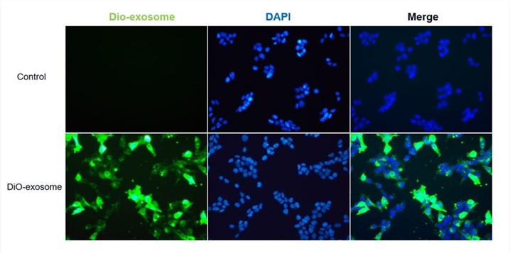 Fig.1 Exosomes labeled were internalized by HEK 293T cells.
Quantitative uptake data provide foundational evidence for understanding how exosomes mediate signaling between donor and recipient cells.
Comparative uptake studies reveal whether certain cell populations display preferential binding or enhanced internalization efficiency.
Uptake quantification strengthens interpretation of downstream changes in proliferation, apoptosis, or differentiation by confirming vesicle entry.
Blocking or modulation experiments can determine whether specific surface molecules contribute to exosome recognition and entry.
For guidance on aligning this assay with your broader research objectives, Creative Biolabs specialists are available.
 Validated Labeling Strategies
Protocols are optimized to preserve exosome morphology and functional integrity while achieving stable signal detection.
Quantitative and Imaging Based Readouts
Both population level and single cell level analyses are available, enabling multidimensional interpretation of uptake behavior.
Flexible Experimental Parameters
Incubation time, concentration ranges, and cell types can be adjusted according to research requirements.
Controlled Background Assessment
Appropriate negative controls and dye only controls are incorporated to minimize misinterpretation caused by nonspecific fluorescence.
Researchers seeking protocol adjustments or specialized cell models may contact Creative Biolabs for technical input.
Understanding how exosomes are internalized by recipient cells is a critical step in interpreting functional outcomes. Through carefully designed labeling strategies and quantitative detection platforms, Creative Biolabs delivers reliable uptake data that strengthen mechanistic studies. For researchers seeking a dependable partner in exosome focused cellular analysis, the Creative Biolabs team is ready to assist with tailored experimental design and technical support.
A: Creative Biolabs incorporates washing procedures, quenching approaches when appropriate, and imaging based confirmation to differentiate surface binding from intracellular localization. Confocal microscopy can be used to validate z axis positioning within cells. Control conditions are included to strengthen interpretation.
A: Yes. Many primary cell types can be accommodated provided they meet viability and culture stability requirements. During project planning, Creative Biolabs evaluates growth conditions and receptor expression considerations to determine suitability and optimize incubation parameters.
A: Comparative designs are feasible. Parallel labeling and standardized incubation conditions can be implemented to allow relative uptake assessment across different exosome preparations. Statistical analysis is structured to support side by side comparison.
A: Common controls include unlabeled exosomes, dye only controls, and receptor blocking conditions when relevant. Creative Biolabs advises on control selection based on the experimental hypothesis to ensure accurate data interpretation.
A: No. This service is intended exclusively for basic and preclinical research. Data are generated to support mechanistic understanding and should not be used for clinical or therapeutic evaluation.