
Autophagy is a tightly controlled intracellular process responsible for the degradation and recycling of cellular components. It plays a central role in maintaining cellular balance, especially under stress conditions such as nutrient deprivation or oxidative challenge. Increasing evidence shows that exosomes can influence autophagic activity by transferring regulatory molecules that affect signaling pathways and lysosomal function.
The exosome modulated autophagy regulation assay service offered by Creative Biolabs supports in vitro studies focused on how exosomes alter autophagy dynamics. By monitoring key markers such as LC3 conversion, autophagosome formation, and lysosomal activity, this assay enables clear evaluation of whether exosomes induce, suppress, or fine tune autophagic flux. Experimental conditions are carefully adjusted based on cell type and research objectives, ensuring that observed changes are biologically meaningful.
| Step | Detail |
|---|---|
| Step 1. Objective Alignment and Model Selection | Creative Biolabs works with researchers to define whether the focus is on autophagy induction, inhibition, or flux modulation, followed by selection of suitable cell systems. |
| Step 2. Culture Preparation and Baseline Stabilization | Cells are cultured under controlled conditions to maintain stable basal autophagy levels prior to treatment. |
| Step 3. Exosome Treatment Configuration | Exosomes are introduced at defined concentrations and time points. Optional conditions such as nutrient deprivation or chemical modulators can be incorporated. |
| Step 4. Autophagy Marker Detection Setup | Appropriate detection strategies are selected, including protein level analysis, fluorescence labeling of autophagic structures, or reporter systems. |
| Step 5. Imaging and Signal Acquisition | Microscopy or biochemical assays are performed to capture autophagosome formation and related changes. |
| Step 6. Flux Assessment and Data Processing | Creative Biolabs evaluates dynamic autophagy changes by comparing treated and control conditions, including flux related measurements where applicable. |
| Step 7. Result Integration and Reporting | Final data include quantitative outputs, representative images, and interpretation aligned with the study objective. |
To refine any stage of the workflow or include additional readouts, Creative Biolabs specialists are available for discussion.
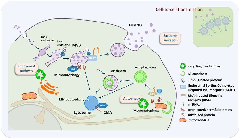 Fig.1 Interaction of Exosome Secretion and Autophagy in Cellular Homeostasis.1
Autophagy assays reveal how exosomes influence degradation pathways and cellular resource management.
Changes in autophagy can reflect the transfer of regulatory molecules that impact signaling pathways.
Autophagy modulation provides insight into how cells adjust to environmental or metabolic stress.
Different exosome preparations can be assessed for their relative ability to regulate autophagic activity.
Researchers aiming to integrate autophagy analysis into broader studies may connect with Creative Biolabs for technical suggestions.
 Multi Marker Detection Capability
Simultaneous evaluation of key autophagy indicators improves interpretation of regulatory effects.
Flux Oriented Analysis Options
Supports assessment of dynamic autophagy activity rather than relying solely on static measurements.
Flexible Experimental Conditions
Allows incorporation of stress models or modulators to better reflect specific research scenarios.
Adaptable to Different Cell Systems
Protocols are adjusted based on cellular characteristics and baseline autophagy levels.
If you need help selecting features that best fit your study, Creative Biolabs scientists are ready to assist.
For comparable support and data quality, getting in touch with Creative Biolabs is a practical next step.
Autophagy regulation provides a critical window into how cells maintain balance and respond to external signals. With carefully designed experiments and reliable detection strategies, Creative Biolabs delivers data that help clarify the role of exosomes in this process. To move forward with your study or discuss customization options, contacting Creative Biolabs is highly recommended.
A: Creative Biolabs evaluates autophagic flux by combining marker analysis with appropriate controls or modulators. This helps determine whether accumulation of autophagosomes reflects activation or impaired degradation.
A: Yes. Conditions such as nutrient limitation or chemical stimulation can be incorporated to assess how exosomes influence autophagy under defined stress scenarios.
A: Common readouts include LC3 conversion, p62 levels, and imaging of autophagic structures. The exact combination depends on the research goal and cell type.
A: Time course studies can be designed to monitor how autophagy evolves after exosome exposure. This approach helps capture early and later responses.
A: Standardized culture conditions, controlled treatment parameters, and replicate analysis are applied to maintain consistency and reliability.
A: Yes. Autophagy data can be aligned with apoptosis, proliferation, or signaling assays to provide a more complete picture of cellular response.
Reference