

The cytoskeleton provides the structural framework that governs cell shape, mechanical stability, and intracellular transport. It also plays a central role in processes such as migration, adhesion, and signal transduction. Exosomes can influence cytoskeletal organization by delivering regulatory molecules that affect actin dynamics, microtubule stability, and associated signaling pathways.
The exosome controlled cytoskeletal organization Assay Service from Creative Biolabs supports in vitro studies focused on how exosomes reshape cytoskeletal architecture and related cellular behavior. By combining imaging based analysis with quantitative assessment of cytoskeletal markers, this assay enables clear evaluation of structural changes following exosome exposure.
| Step | Detail |
|---|---|
| Step 1. Experimental Objective Definition and Cell Model Selection | Creative Biolabs works with researchers to clarify whether the study targets structural rearrangement, signaling related changes, or functional outcomes, followed by selection of appropriate cell systems. |
| Step 2. Culture Preparation and Baseline Stabilization | Cells are maintained under controlled conditions to establish consistent cytoskeletal organization before treatment. |
| Step 3. Exosome Treatment Planning | Exosomes are applied at defined concentrations and incubation periods, with timing selected to capture early structural responses or longer term reorganization. |
| Step 4. Cytoskeletal Staining and Labeling | Cells are processed using validated staining methods targeting actin filaments, microtubules, or associated proteins. |
| Step 5. Imaging Acquisition | High resolution imaging is performed to visualize cytoskeletal structures, including filament distribution and organization patterns. |
| Step 6. Quantitative Analysis | Creative Biolabs evaluates parameters such as filament density, alignment, and structural integrity across treatment groups. |
| Step 7. Data Interpretation and Reporting | Results are normalized and presented with both visual and numerical outputs to support clear comparison. |
| Step 8. Final Data Delivery | Reports include raw images, processed data, and methodological details for further analysis. |
To customize workflow steps or incorporate additional markers, you can connect with our specialists for further discussion.
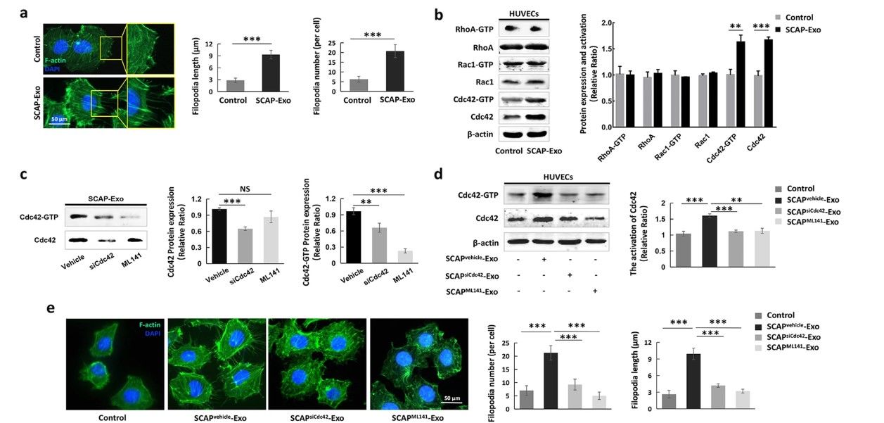 Fig.1 SCAP-Exo enhanced HUVEC migration through Cdc42-driven cytoskeletal changes.1
Cytoskeletal organization reflects how cells maintain shape and respond to external signals.
Changes in filament arrangement can indicate altered cellular stiffness and dynamics.
Comparative studies reveal how different exosome preparations influence structural behavior.
Cytoskeletal data provide a foundation for interpreting movement and cell environment interactions.
Researchers interested in incorporating cytoskeletal analysis into their studies can reach out to Creative Biolabs for technical input.
 Detailed Structural Visualization
High quality imaging enables precise observation of cytoskeletal arrangement and organization.
Quantitative Morphological Analysis
Provides measurable parameters that support objective comparison between conditions.
Flexible Marker Selection
Supports targeting of actin, microtubules, and associated regulatory proteins.
Integration with Functional Assays
Cytoskeletal data can be correlated with migration, adhesion, or signaling studies.
If you need assistance in selecting suitable features, Creative Biolabs scientists are ready to help.
For similar project support, reaching out to Creative Biolabs is a practical step.
Cytoskeletal organization is central to how cells maintain structure and respond to external cues. With carefully designed experiments and reliable analytical methods, Creative Biolabs delivers data that support detailed in vitro research on exosome driven structural changes. For customized assay development or to begin your study, contacting Creative Biolabs is strongly encouraged.
A: The assay can include analysis of actin filaments, microtubules, and associated proteins. Marker selection is based on the research objective and cell model.
A: Yes. High resolution imaging combined with quantitative analysis allows detection of both major and subtle changes in cytoskeletal organization.
A: Time course experiments can be designed to observe how cytoskeletal structures change over time following exosome exposure.
A: Standardized staining protocols, controlled imaging conditions, and replicate analysis are applied to maintain reproducibility.
A: Yes. Structural changes can be correlated with migration, adhesion, or signaling assays to provide deeper insight into cellular behavior.
A: Factors such as cell density, culture conditions, and staining quality can affect results. Creative Biolabs carefully controls these parameters to ensure reliable data.
Reference