Introduction What Can We Offer? Workflow Published Data Why Choose Us? Applications FAQs Featured Services Featured Products
Creative Biolabs has successfully constructed a series of improved and innovative aptamer platforms to provide fast and better 3D structure determination services for our worldwide customers. Our high-quality services can help you get satisfactory results without repeated redundant trials and obtain a milestone development in your aptamer development.
Contact our team to get an inquiry now!
Aptamer 3D Structure Determination
Aptamers are single-stranded oligonucleotides that function as "chemical antibodies" by folding into unique three-dimensional shapes. While primary sequences are easily obtained via SELEX, it is the tertiary structure, comprising hairpins, G-quadruplexes, and pseudoknots, that dictates target recognition. Modern research emphasizes that structural determination is no longer optional; it is the cornerstone of rational design. By resolving these structures at atomic resolution, we enable the visualization of hydrophobic interactions, base stacking, and induced-fit mechanisms that define binding affinity and specificity.
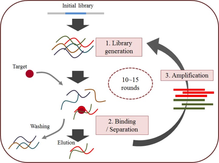 Fig.1 The general SELEX strategy.1
What Can We Offer?
Creative Biolabs offers a comprehensive suite of experimental and computational services to define the 3D topology of aptamers and their target complexes. Our platform bridges the gap between theoretical modeling and empirical validation, ensuring a holistic understanding of molecular function.
Our Core Service Capabilities:
High-Resolution NMR Spectroscopy
We utilize heteronuclear multidimensional NMR to study aptamers in their native solution state. This is ideal for sequences under 50 nt, providing data on conformational dynamics and ligand-induced folding changes.
X-Ray Crystallography Services
For atomic-level blueprints, we provide full crystallization screening of aptamer-target complexes. We resolve diffraction patterns to provide the exact spatial coordinates of every atom within the binding pocket.
Cryo-EM & Electron Crystallography
We leverage state-of-the-art Cryo-Electron Microscopy to characterize large aptamer-protein assemblies. This "native-state" imaging avoids the artifacts of crystallization and is perfect for membrane-bound targets.
Small-Angle X-ray Scattering (SAXS)
We offer SAXS to determine the low-resolution global shape and flexibility of aptamers in bulk solution, ensuring the 3D models align with physiological behavior.
Workflow
01
Initial Technical Consultation: We discuss your aptamer sequence, target properties, and resolution requirements.
02
Sample Provision: Clients provide purified aptamer sequences (lyophilized or in buffer) and, if applicable, the target protein/molecule.
03
Standardized Stabilization: We perform thermal conditioning (95℃ for 5 minutes followed by controlled cooling) to ensure homogeneous folding.
04
Data Acquisition: Samples undergo NMR, X-ray, or Cryo-EM analysis based on the selected strategy.
05
Structure Refinement: Our bioinformaticians process raw data into a refined 3D PDB model.
06
Final Delivery: You receive a comprehensive scientific report, the 3D atomic model (PDB format), and high-resolution density maps or spectra.
Published Data
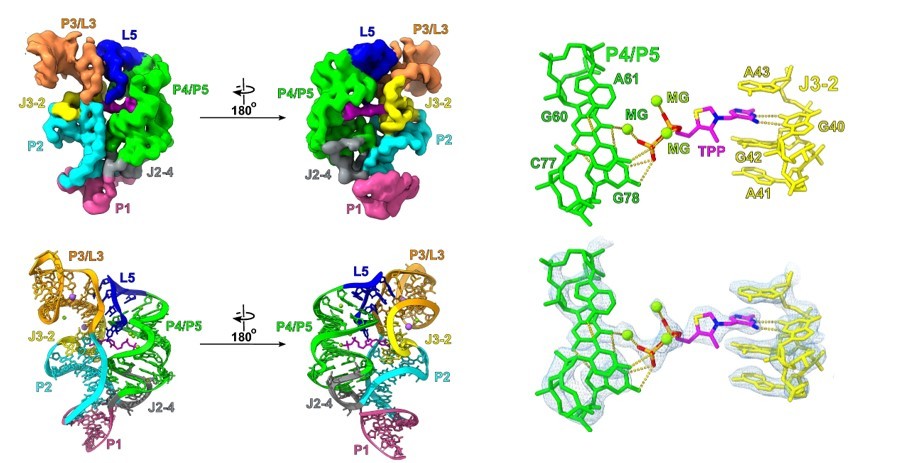 Fig.2 The cryo-EM structure of the TPP riboswitch.2
In this study, researchers demonstrated a novel strategy for high-resolution of small RNAs by fusing them to a group II intron scaffold. This approach was utilized to resolve the structure of the 86-nucleotide thiamine pyrophosphate (TPP) riboswitch aptamer domain, a historically difficult target due to its small molecular weight. The project results achieved a remarkable 2.5 Å resolution of the TPP riboswitch, providing unprecedented visualization of the ligand-binding pocket. Furthermore, by determining the structure of the ligand-free "apo" state, the researchers observed that the aptamer adopts an open Y-shaped conformation in the absence of its ligand. This case study confirms that scaffolding technologies can bypass the resolution limits of traditional cryo-EM for small aptamers, enabling the capture of multiple conformational states and facilitating more precise ligand-affinity engineering.
Why Choose Us?
-
Advanced Technological Integration: We combine NMR, X-ray, and Cryo-EM to provide multi-angle structural validation.
-
Standardized Stabilization Protocols: Our proprietary cooling methods ensure structural homogeneity across all samples.
-
Atomic-Level Precision: We focus on resolving specific interactions like π-stacking and hydrogen bonding.
-
Extensive Aptamer Expertise: Our team has years of experience specifically in oligonucleotide folding.
-
Rapid Turnaround Times: Integrated workflows allow for faster delivery of PDB models compared to academic labs.
-
End-to-End Support: From initial SELEX consultation to final structural optimization, we support the entire development lifecycle.
Applications
-
Rational Sequence Truncation: By identifying non-essential loops and tails in the 3D model, we help clients reduce 80-mer pools into 20-mer leads, lowering synthesis costs and improving tissue penetration. This structural streamlining is vital for enhancing the bio-distribution of therapeutic oligonucleotides.
-
Mechanism of Action (MoA) Elucidation: We visualize exactly how an aptamer inhibits its target, whether through competitive site blocking or allosteric modulation, providing essential data for regulatory filings and precise functional characterization that explains the biological impact of the molecule.
-
Structure-Based Optimization: Atomic models allow for the rational insertion of chemical modifications (e.g., 2'-Fluoro or 2'-OMe) at specific positions to enhance biostability and pharmacokinetic profiles without disrupting the binding pocket's architecture or the essential target-ligand orientation.
-
Advanced Diagnostic and Delivery Engineering: Accurate tertiary models enable the precise conjugation of fluorophores or drug payloads at positions that do not interfere with binding, optimizing aptamers for molecular imaging and targeted delivery for highly specific diagnostic applications.
FAQs
Q: How do you ensure the aptamer isn't forming multiple different shapes in the sample?
A: We use a standardized thermal denaturation and slow-refolding protocol, followed by Dynamic Light Scattering (DLS) and analytical SEC to verify the monodispersity and structural homogeneity of the final sample before data collection.
Q: Can you determine the structure of an aptamer bound to a small molecule?
A: Yes, we utilize high-resolution NMR and X-ray crystallography to resolve aptamer-small molecule complexes, which is vital for biosensor development and metabolite tracking in complex physiological matrices where high sensitivity is paramount.
Q: Is it possible to resolve the structure of very long aptamers (over 100 nt)?
A: Large aptamers are challenging for NMR; however, we utilize Cryo-EM or "divide and conquer" NMR strategies where we resolve fragments independently and reconstruct the whole model using advanced computational merging and refinement.
Q: Does your service include docking simulations?
A: Yes, we provide integrated molecular docking and MD simulations to complement experimental data, helping to visualize the energetic stability and binding kinetics of the resulting complex under varied physiological conditions.
Q: Can 3D structure determination help if my aptamer has poor stability in serum?
A: Yes, by identifying flexible regions prone to nuclease attack in the 3D model, we can suggest specific chemical modifications or structural reinforcements to stabilize those areas while maintaining optimal target binding affinity.
Q: How do you account for structural dynamics or multiple conformational states?
A: We employ ensemble modeling and molecular dynamics to capture flexibility. Our cryo-EM workflows also utilize 3D classification to resolve heterogeneous populations, ensuring your final model reflects the most biologically relevant state under varying temperature and buffer conditions.
Creative Biolabs stands as a leader in high-resolution aptamer analysis, combining decades of biological expertise with cutting-edge structural platforms. If you are looking to unlock the full potential of your sequences, we invite you to contact our specialists for a detailed project assessment.
Featured Services
Featured Products
References
-
Song, Kyung-Mi, Seonghwan Lee, and Changill Ban. "Aptamers and their biological applications." Sensors 12.1 (2012): 612-631. Distributed under Open Access license CC BY 3.0, without modification. https://doi.org/10.3390/s120100612
-
Haack, Daniel B., et al. "Scaffold-enabled high-resolution cryo-EM structure determination of RNA." Nature Communications 16.1 (2025): 880. Distributed under Open Access license CC BY 4.0, without modification. https://doi.org/10.1038/s41467-024-55699-5
For Research Use Only.
Related Sections: