Introduction What Can We Offer? Workflow Published Data Why Choose Us? Applications FAQs Featured Services Featured Products
Creative Biolabs provides specialized 3D structure modeling services to decode the spatial complexities of nucleic acid ligands. We help researchers move beyond simple sequence data, solving critical challenges in binding mechanism elucidation and structural stability to accelerate high-performance aptamer development.
Contact our team to get an inquiry now!
Aptamer 3D Structure Modeling
Aptamers function as "chemical antibodies" whose efficacy is dictated by their sophisticated tertiary folds, such as G-quadruplexes, pseudoknots, and intricate stem-loop clusters. 3D structure modeling is the essential bridge between primary sequence discovery and functional optimization. Recent research highlights that even minor sequence variations can lead to dramatic shifts in the spatial orientation of binding loops. By utilizing computational techniques to simulate these folds, we provide a high-resolution map of the aptamer's architecture. This spatial understanding is critical for identifying active binding sites, predicting environmental stability, and guiding the rational design of modifications that enhance affinity and specificity in complex biological matrices.
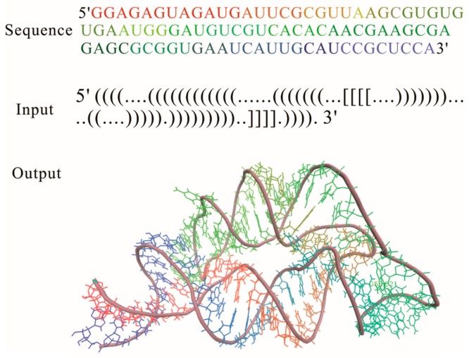 Fig.1 3D structure for the THF riboswitch aptamer.1,2
What Can We Offer?
Creative Biolabs provides a comprehensive suite of computational modeling and simulation services designed to reveal the hidden geometry of your aptamers. We offer end-to-end structural characterization, from initial folding predictions to complex target-docking simulations, ensuring a robust biophysical profile for every lead.
Our Core Modeling Services:
Template-Based Homology Modeling
We utilize advanced algorithms to align your sequences with known structural motifs in the Protein Data Bank (PDB). By identifying conserved domains, we build highly accurate 3D models based on established structural templates.
Ab Initio Folding
Simulations
For novel or synthetic sequences without known analogs, we employ physics-based methods like Fragment Assembly of RNA (FARFAR). We simulate thousands of possible conformations to identify the global minimum free energy state.
Molecular Dynamics (MD) Simulations
Our team performs all-atom MD simulations in various solvent environments. We observe the structural fluctuations of your aptamer over time to ensure the fold remains stable under physiological ion concentrations and temperatures.
Aptamer-Target Docking Analysis
We predict the precise interaction interface between your aptamer and its target protein or small molecule. By calculating binding energies and identifying contact residues, we provide a blueprint for enhancing molecular recognition.
Rational Truncation & Optimization
Using the 3D model as a guide, we identify non-essential "scaffolding" nucleotides. This allows us to recommend truncated versions that maintain or improve affinity while significantly reducing chemical synthesis costs.
Workflow
01
Initial Consultation: We discuss your target specifications, SELEX conditions, and project goals.
02
Material Submission: Clients provide the aptamer sequence (DNA or RNA) and, if applicable, the target protein sequence or structure.
03
Secondary Structure Prediction: We establish the 2D landscape (mfold/RNAfold) to identify baseline stem-loop regions.
04
3D Tertiary Assembly: Using hybrid template/ab initio methods, we construct the spatial model.
05
Refinement & Simulation: We perform energy minimization and MD simulations to validate structural integrity.
06
Binding Analysis: Docking simulations are conducted to visualize the aptamer-target complex.
07
Final Delivery: You receive a comprehensive report containing PDB files, interaction maps, and optimization recommendations.
Published Data
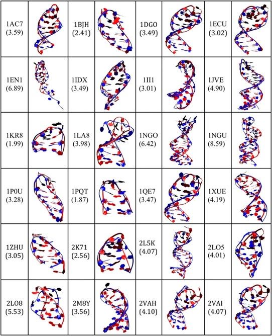 Fig.2 3D predicted structures (ssDNA colored red) of the 24 ssDNA hairpin structures.2,3
In this study, researchers established a sophisticated pipeline for predicting the three-dimensional structures of single-stranded (ss) DNA hairpins, which are fundamental motifs in aptamer-based biosensors. By integrating secondary structure prediction with 3D ssRNA modeling and subsequent transformation into refined ssDNA 3D structures. The project demonstrated that 3D modeling accurately identifies critical binding domains, allowing for the rational truncation of sequences while maintaining or enhancing target affinity. This high-resolution approach provides a robust framework for improving the consistency and reproducibility of aptamer-based devices, turning computational insights into validated molecular tools.
Why Choose Us?
-
Expertise in Nucleic Acid Physics: Deep specialization in the unique folding rules of DNA and RNA.
-
Advanced Computational Power: High-performance clusters capable of running long-range MD simulations.
-
Data-Driven Optimization: We don't just provide a model; we provide actionable strategies for truncation.
-
Transparent Methodology: Every simulation parameter is documented, ensuring reproducible and publishable results.
-
Rapid Turnaround: Optimized workflows allow us to deliver high-resolution models in a fraction of the time of NMR.
-
Integrated Solutions: Seamless transition from structural modeling to chemical synthesis and assay validation.
Applications
-
Therapeutic Lead Optimization: This process identifies the minimum functional core of an aptamer to enhance tissue penetration while reducing synthesis costs. Modeling of structural hotspots facilitates the rational design of therapeutic agents with enhanced pharmacokinetic profiles and superior binding kinetics.
-
Diagnostic Probe Design: Precise modeling ensures that binding loops remain accessible and active when conjugated to fluorophores, gold nanoparticles, or electrochemical surfaces. Geometric analysis prevents steric hindrance and unintended structural collapse, which are common hurdles in high-sensitivity sensor development.
-
Environmental Biosensor Engineering: Structural stability and folding resilience are evaluated across diverse pH levels, salinities, and temperatures for field-deployable tools. This ensures the engineering of robust aptamers capable of maintaining consistent molecular recognition under harsh, non-physiological conditions.
-
In Silico Cross-Reactivity Screening: This application predicts potential off-target binding to structural homologs or non-target proteins. Computational pre-screening reduces experimental overhead, identifies off-target motifs early in development, and significantly improves the safety profiles of molecular ligands.
-
Payload Conjugation Strategies: Modeling guides the precise attachment of siRNAs, toxins, or imaging agents at positions that do not disturb the tertiary fold. These simulations ensure that the active binding domain remains undisturbed and functional during the assembly of complex drug conjugates.
FAQs
Q: What is the primary difference between your homology modeling and ab initio services?
A: Homology modeling relies on existing structural templates in databases, making it faster and highly accurate for known motifs. In contrast, ab initio modeling builds the structure from first principles using physical laws, which is indispensable for entirely novel sequences where no structural data exists.
Q: Can you model aptamers with chemical modifications like 2'-F or 2'-OMe?
A: Yes, our advanced force fields are specifically calibrated to handle common base and backbone modifications. We can predict with high precision how these changes affect both the global tertiary fold and the specific local binding interactions.
Q: How do you validate the accuracy of a predicted 3D model?
A: We employ a multi-step validation process using energy profile analysis, Ramachandran-like plots for nucleic acids, and comparison with known experimental data. We also offer extended MD simulations to confirm the model reaches a stable, reproducible equilibrium.
Q: What information do I need to provide for a docking study?
A: We require the complete aptamer sequence and either the PDB ID of the target or its amino acid sequence. If the target structure is unknown, we provide protein homology modeling as a complementary service to facilitate the docking.
Q: Can structural modeling help if my aptamer has poor serum stability?
A: Yes. By identifying "exposed" loops that are particularly vulnerable to nuclease degradation, we can suggest internal modifications, structural tightening, or protective capping strategies to significantly improve the half-life of your candidate.
Q: Do you offer assistance in interpreting the final docking results?
A: Our senior biologists provide a comprehensive narrative report that explains the chemical significance of the interactions, including hydrogen bonding, pi-stacking, and electrostatic forces, to help guide your subsequent experimental validation.
Q: Can modeling predict the effect of temperature on binding?
A: Through temperature-scaled MD simulations, we can estimate the melting profile of the aptamer structure and predict binding stability at various thermal setpoints, ensuring performance across a range of operational environments.
Creative Biolabs combines decades of biological expertise with cutting-edge computational tools to deliver unmatched structural insights. Contact our team today to discuss your project requirements and discover how our modeling services can elevate your aptamer research to the next level.
Featured Services
Featured Products
References
-
Gong, Sha, et al. "Computational methods for modeling aptamers and designing riboswitches." International journal of molecular sciences 18.11 (2017): 2442. https://doi.org/10.3390/ijms18112442
-
Jeddi, Iman, and Leonor Saiz. "Three-dimensional modeling of single stranded DNA hairpins for aptamer-based biosensors." Scientific Reports 7.1 (2017): 1178. https://doi.org/10.1038/s41598-017-01348-5
-
Distributed under Open Access license CC BY 4.0, without modification.
For Research Use Only.
Related Sections: