Introduction What We Can Offer Workflow Published Data Why Choose Us? Applications FAQs Featured Services Featured Products
Analyzing the structure of your interest aptamers and their binding complex is beneficial for the exploration of potential therapeutic aptamers and targets. Creative Biolabs is a leading biotechnology company that has the expertise and ability to provide aptamer structure analysis services for customers worldwide.
Contact our team to get an inquiry now!
Aptamer and Aptamer Structure Analysis
Aptamers, or "chemical antibodies," are single-stranded oligonucleotides that fold into intricate three-dimensional shapes to bind targets with high specificity. Unlike traditional linear sequences, their function is dictated by spatial conformations such as G-quadruplexes, pseudoknots, and hairpins. Modern research, including 2025 structural surveys, reveals that nearly 25% of DNA aptamers possess "hidden" quadruplex motifs that govern binding efficacy. Aptamer structure analysis is the process of deciphering these 3D blueprints and the thermodynamics of "induced-fit" mechanisms. Understanding these interactions is essential for rational design, allowing for precise truncation and chemical modification to maximize biostability and affinity.
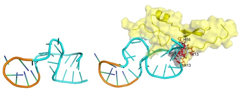 Fig.1 Schematic diagram of aptamer conformational recognition of targets to form an aptamer-target complex.1,3
What We Can Offer
We provide a comprehensive analytical suite designed to map the structural landscape of your aptamers at every level, from initial secondary topology to high-resolution atomic coordinates of the aptamer-target complex.
Our Specialized Services include:
2D Secondary Structure Mapping
We utilize chemical probing (SHAPE/DMS) and enzymatic digestion to establish a foundational map of paired and unpaired regions. This ensures that the computational models for your leads are grounded in empirical biochemical data.
3D Structure Determination via NMR
Our high-field NMR spectroscopy is the gold standard for studying aptamers in their native solution state. We perform imino proton fingerprinting to confirm base-pairing and utilize NMR titrations to visualize exactly how your aptamer shifts conformation upon binding its target.
High-Resolution X-Ray Crystallography
For projects requiring atomic-level precision, we crystallize aptamer-target complexes. This allows us to identify the specific hydrogen bonds and van der Waals forces driving the interaction, providing the ultimate "molecular fingerprint" for IP protection.
Cryo-EM for Large Complexes
We leverage Cryo-Electron Microscopy, enhanced by RNA Origami scaffolds, to resolve the structures of aptamers bound to large or difficult targets, such as membrane proteins and viral particles.
Advanced 3D Modeling & AI Integration
Using the latest databases and AI tools, we provide predictive modeling and molecular dynamics simulations to prioritize sequences for synthesis and experimental validation.
G-Quadruplex & i-Motif Profiling
We specifically screen G-rich sequences for quadruplex propensity, evaluating their stability in various ionic environments (K+ vs. Na+) to ensure predictable performance in physiological settings.
Workflow
01
Technical Consultation: We discuss your target type, known sequence data, and specific project goals to select the optimal analytical approach.
02
Material Provision: Clients provide the aptamer sequence or synthesized material and the purified target molecule (protein, small molecule, or cell fragment).
03
Sequence Verification & 2D Probing: We begin with SHAPE or DMS probing to establish the secondary structure constraints.
04
Experimental Data Acquisition: Depending on the chosen path, we perform NMR, X-ray diffraction, or Cryo-EM imaging.
05
Thermodynamic Correlation: We integrate structural data with ITC (Isothermal Titration Calorimetry) to map the energetic landscape of the binding event.
06
Final Delivery: You receive a comprehensive structural report, including high-resolution 3D models (PDB format), interaction maps, and optimization recommendations.
Published Data
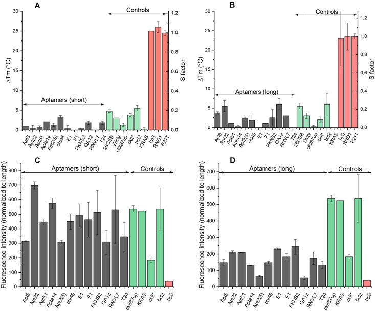 Fig.2 Experimental validation of G4s formation.2,3
In this comprehensive 2025 study, researchers analyzed over 1,400 aptamer sequences to investigate their propensity for forming G-quadruplex (G4) structures. The research revealed that nearly 25% of DNA aptamers and 16% of RNA aptamers are predicted to form stable G-quadruplex folds, yet the word "quadruplex" appeared in only 17% of the original reporting articles. By experimentally testing 30 candidate sequences using circular dichroism and ThT fluorescence, the team confirmed G4 formation in all sequences with a G4Hunter score above 1.31. These findings highlight a significant "structural blind spot" in aptamer development, demonstrating that G4 motifs often serve as a universal recognition core that remains undetected by standard sequence analysis. Leveraging these insights, it is possible to identify and characterize these hidden structural modules, ensuring superior binding specificity and stability for their leads.
Why Choose Us?
-
Multi-Platform Technical Suite: We offer a unique combination of NMR, X-ray, Cryo-EM, and chemical probing for total structural clarity.
-
Industry-Leading AI Integration: Our workflows incorporate the latest structure-function databases for AI-ready data generation.
-
Customized Optimization Strategies: We don't just provide data; we provide a roadmap for truncation and chemical modification.
-
Rigorous Quality Control: Every structural model is validated against empirical biochemical constraints to ensure accuracy.
-
Direct Expert Access: You work directly with PhD-level structural biologists from initial consultation through final delivery.
Applications
-
Precision Drug Design & Truncation: Identifying the minimum essential binding motif allows for the removal of non-functional nucleotides, facilitating the synthesis of compact, high-performance therapeutic leads with improved tissue penetration.
-
Biostability Enhancement via Rational Modification: Mapping the folding topology enables the targeted introduction of 2'-O-methyl, 2'-fluoro, or phosphorothioate modifications at non-interfering sites to maximize nuclease resistance while maintaining the binding pocket's integrity.
-
Intellectual Property Protection: High-resolution 3D structural data provides a "molecular fingerprint" that clearly defines the unique mechanism of action, significantly strengthening patent claims and providing a scientific barrier against competitor sequences.
-
Advanced Biosensor Engineering: Structural insights are vital for developing "signal-on" biosensors, where target binding must trigger a specific, predictable conformational switch or displacement event to generate a measurable optical or electrochemical signal.
-
Off-Target Risk Assessment & Selectivity Mapping: By identifying common structural motifs, such as G-quadruplexes or specific stem-loops, researchers can proactively predict and mitigate cross-reactivity with endogenous proteins, reducing safety risks during subsequent development phases.
-
Molecular Dynamics & Interaction Modeling: Detailed atomic structures serve as the foundation for computational simulations, allowing us to predict how environmental changes like pH or temperature influence the aptamer's thermodynamic stability and target affinity.
FAQs
Q: Can you resolve structures of aptamers bound to small-molecule ligands?
A: Yes, we utilize a combination of NMR imino proton shifts and X-ray crystallography to map the binding pockets of small-molecule aptamers, including those with sub-nanomolar affinities.
Q: Why is G-quadruplex analysis important if my sequence wasn't designed to form one?
A: Many G-rich sequences spontaneously adopt quadruplex folds during the SELEX process. Identifying these is crucial because they often dictate the stability and specificity of the aptamer in different ionic environments.
Q: Do you offer services for aptamers containing unnatural or modified bases?
A: Our team is highly experienced in analyzing modified oligonucleotides. We adjust our NMR and modeling parameters to account for the unique geometry and electronic properties of 2'-modified or backbone-altered bases.
Q: Is it possible to analyze aptamers that bind to membrane-bound proteins?
A: We utilize Cryo-EM in conjunction with nanodisc technology or detergent micelles to resolve structures of aptamers interacting with membrane-embedded targets in a near-native environment.
Q: How does structural analysis help in reducing the cost of aptamer production?
A: By identifying the "minimal binding domain," we can often truncate the aptamer sequence by 30-50%. Shorter sequences are significantly cheaper to synthesize at a commercial scale.
Q: Can you help if my aptamer has multiple folding states (conformational heterogeneity)?
A: This is a common challenge that we address using dynamic NMR and SAXS (Small-Angle X-ray Scattering) to characterize the ensemble of states and identify conditions that favor the active conformation.
Q: What happens if the aptamer fails to crystallize during the project?
A: In such cases, we pivot to solution-state NMR or Cryo-EM. Our multi-tiered approach ensures that we can still provide high-quality structural insights even when traditional methods face hurdles.
Creative Biolabs' expert structural biologists are dedicated to transforming your aptamer sequences into high-performance molecular tools. Our comprehensive biophysical insights ensure that your leads are optimized for maximum efficacy and stability. Contact us today to discuss your project and receive a tailored structural analysis plan.
Featured Services
Featured Products
References
-
Zhang, Ning, et al. "Structural biology for the molecular insight between aptamers and target proteins." International Journal of Molecular Sciences 22.8 (2021): 4093. https://doi.org/10.3390/ijms22084093
-
Cucchiarini, Anne, et al. "Analysis of quadruplex propensity of aptamer sequences." Nucleic Acids Research 53.9 (2025): gkaf424. https://doi.org/10.1093/nar/gkaf424
-
Distributed under Open Access license CC BY 4.0, without modification.
For Research Use Only.
Related Sections: