Studies have shown that different from erythrocytes, several complement channels are required for the lysis of a nucleated mammalian cell. The propidium iodide (PI), which belongs to phenanthridine intercalators, is highly polar and water soluble to exclude reagents from passing through the cell membrane of living cells. When PI enters the cell, it will be tightly combined with the nucleus by being embedded in the DNA. Furthermore, the excitation and emission wavelengths of PI allow it to be detected by fluorescence microscopy replication or flow cytometry. Here, Creative Biolabs has developed the novel propidium iodide assay protocol to promote your research. Please note that our protocols are only for your reference.
Propidium Iodide Assay Protocol
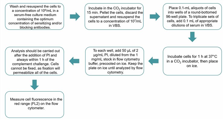 Fig.1 Flow chart of the propidium iodide assay protocol. (Creative Biolabs)
Creative Biolabs has long-lasting expertise in the field of the human complement system. Our team of experts can assist you in designing the best study outline customized to meet the requirements of your discovery/development program. We value our client’s feedback and are committed to striving for one hundred percent client satisfaction. For customers' special project needs, we can also provide customized services based on standard protocols.
Published Data
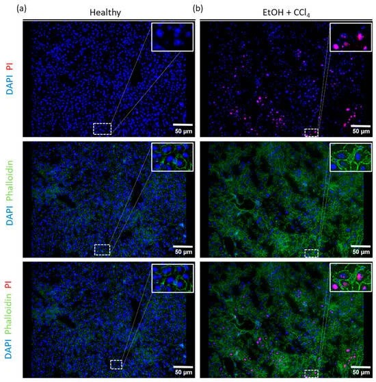 Fig 2. Co-staining of phalloidin and PI.1
Research shows that propidium iodide (PI) is a reliable marker for detecting dead or dying cells in frozen liver tissue sections. Compared to the traditional immunohistochemical TUNEL staining method, PI staining is rapid, simple, and provides more qualitative information along with economic advantages. PI staining serves as a valuable technique for detecting dead or dying cells within particular regions of tissue. This method enhances our understanding of the disease’s pathobiology and the interactions between cells that happen in proximity to areas of cell death. For instance, in liver tissue, identifying PI-positive cells can highlight regions of tissue damage or stress that align with specific disease-related zoning patterns. Additionally, when used in multiplex immunofluorescence methods, PI staining can detect immune cells in the surrounding tissue microenvironment simultaneously, which aids in understanding local immune responses.
Services and Products at Creative Biolabs
If you are interested in our products and services, please do not hesitate to contact us for more information.
Reference
-
Krapoth, Tim Christopher, et al. "Wanted: Dead or Alive Cells with Propidium Iodide Staining in Liver Tissue." International Journal of Molecular Sciences 25.24 (2024): 13521. Distributed under Open Access license CC BY 4.0, without modification.
For Research Use Only.
Related Sections: