Immunophenotyping based T Cell Exhaustion Service
Background Service Highlights FAQs Contact Us
Introduction: Deciphering T-Cell Dysfunction in Modern Therapeutics
T-cell exhaustion is a state of profound cellular dysfunction that arises during chronic antigen exposure, representing a significant barrier to effective immunity in oncology and infectious disease. This dysfunctional state is not a simple "off-switch" but a distinct differentiation pathway leading to the hierarchical loss of effector functions, including proliferative capacity and cytotoxic potential. Exhausted T cells are defined by the sustained co-expression of multiple inhibitory receptors, often called immune checkpoints, and a distinct transcriptional and epigenetic landscape. Understanding this complex cellular state is paramount for the development of next-generation immunotherapies. At Creative Biolabs, we recognize that accurately quantifying the gradient of T-cell exhaustion within the tumor microenvironment (TME) or during chronic infection is critical for predicting therapeutic response and elucidating mechanisms of resistance to checkpoint blockade. Our services provide the granular, single-cell resolution necessary to dissect these intricate immune dynamics.
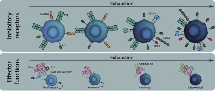 Fig.1 Characteristics of exhausted T cells.1
Our Immunophenotyping based T Cell Exhaustion Service
Creative Biolabs provides a comprehensive, high-dimensional immunophenotyping service engineered to deliver an unparalleled analysis of T-cell exhaustion. This service is designed to support academic, biotechnology, and pharmaceutical partners in advancing their immuno-oncology and infectious disease programs. By leveraging state-of-the-art multi-color flow cytometry, we move beyond simplistic marker analysis to build a detailed, quantitative map of the immune landscape. The primary objective is to characterize the precise phenotype and functional capacity of T-cell subsets, enabling the identification of distinct exhaustion states, from early dysfunction to terminal exhaustion. This level of detail is critical for evaluating the pharmacodynamic effects of novel checkpoint inhibitors, characterizing CAR-T cell products, and identifying patient populations most likely to benefit from immunotherapeutic intervention.
Service Contents
Our analytical workflow is built on a foundation of meticulously designed and validated multi-color flow cytometry panels that provide a holistic view of T-cell exhaustion.
Comprehensive Inhibitory Receptor Profiling
The core of our service is the precise quantification of co-inhibitory receptor expression on individual CD4+ and CD8+ T cells. Our standard and customizable panels include, but are not limited to, the canonical markers of exhaustion:
-
PD-1 (CD279): A foundational marker for identifying exhausted populations.
-
TIM-3 (CD366): Critical for identifying terminally differentiated, severely dysfunctional T cells, often co-expressed with PD-1.
-
LAG-3 (CD223): A key checkpoint that contributes to T-cell dysfunction, particularly in the TME.
-
CTLA-4 (CD152): An essential checkpoint for regulating T-cell activation and tolerance.
-
TIGIT, CD160, and 2B4 (CD244): Additional receptors that allow for the delineation of intermediate and distinct subsets of exhausted cells.
Functional Status Assessment via Intracellular Cytokine Staining (ICS)
A phenotypic profile is incomplete without a functional correlate. Following in vitro stimulation, we employ ICS to measure the single-cell production of key effector cytokines. This reveals the functional capacity of specific T-cell subsets and confirms the loss of function characteristic of exhaustion. Key analytes include:
-
Interferon-gamma (IFN-γ): A critical cytokine for anti-viral and anti-tumor immunity.
-
Tumor Necrosis Factor-alpha (TNF-α): A pro-inflammatory cytokine indicative of a potent effector response.
-
Interleukin-2 (IL-2): A cytokine essential for T-cell proliferation, the production of which is one of a T cell’s first functions to be lost during the progression to exhaustion.
Flexible Sample Input and Processing
We have optimized protocols for a wide range of biological materials:
-
Peripheral Blood (Whole Blood or PBMCs)
-
Bone Marrow Aspirates
-
Dissociated Primary Tissues (e.g., tumor, lymph node)
-
Cultured Cell Lines
A key Creative Biolabs innovation is our whole-blood staining protocol, which eliminates the need for density gradient separation. This reduces sample manipulation artifacts, minimizes processing time, and critically, retains the complete leukocyte profile, including vital granulocyte populations often lost during PBMC isolation.
High-Dimensional Data Analysis and Gating Strategy
Data is acquired on advanced cytometers capable of resolving more than 20 parameters simultaneously. Our bioinformaticians employ sophisticated, hierarchical gating strategies to isolate specific cell populations with precision. This allows for the detailed analysis of exhaustion marker expression within memory and effector T-cell compartments, providing a complete picture of the immune response.
Our Advantages
Choosing Creative Biolabs provides access to over two decades of industry expertise in cellular immunology, translated into a service defined by scientific rigor and technological superiority.
-
High-Resolution Multi-Color Flow Cytometry
-
Expert-Designed Panels and Validated Protocols
-
High-Throughput Capabilities
-
Integrated Cytokine and Phenotype Analysis
FAQs
Q1: How do you distinguish between T-cell exhaustion and anergy?
A1: This distinction is made through multi-parameter analysis. While both are dysfunctional states, they possess distinct phenotypic and functional profiles. Exhaustion is characterized by the high and sustained co-expression of multiple inhibitory receptors like PD-1 and TIM-3 and a progressive loss of function, whereas anergy can be reversible and has a different surface marker signature.
Q2: Can the analysis panel be customized?
A2: Yes. While we offer expertly validated core panels, we specialize in collaborating with our clients to design and validate custom panels that incorporate specific markers of interest relevant to your unique therapeutic modality or disease model.
Q3: What data deliverables can I expect?
A3: Our comprehensive data package includes raw and compensated flow cytometry data files, detailed gating strategies, and a full report summarizing the percentage and expression levels (MFI) of all markers on specified T-cell subsets, presented with publication-quality figures and statistical analysis.
Contact Us
To advance your therapeutic program with unparalleled insight into T-cell exhaustion, partner with Creative Biolabs. Our team of specialists is ready to discuss how our advanced immunophenotyping services can be tailored to meet your specific research and development objectives.
Reference
-
Jenkins, Edward et al. "The current state and future of T-cell exhaustion research." Oxford open immunology vol. 4,1 iqad006. 8 Jul. 2023. DOI: 10.1093/oxfimm/iqad006. Distributed under Open Access License CC BY 4.0, without modification.
 Download our brochure