Background Services Overview Published Data Protocols Related Products Q&A Resources
Of all the autoantibodies that target complement proteins, anti-C1q autoantibodies have received the most attention, and anti-C1q autoantibody tests have become particularly important. Creative Biolabs has solid expertise in the field of complement autoantibodies, which has prompted us to provide our customers with the best autoantibody testing services. Our professional team will work shoulder to shoulder with our customers to meet their research needs.
Background
Overview of Anti-C1q Autoantibody
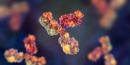
Complement C1q is a cationic glycoprotein produced by immune cells (macrophages, monocytes, and dendritic cells), fibroblasts, and epithelial cells. It is the first component identified in the activation cascade of the classical complement pathway, which functions mainly to clear autoantigens generated during cell apoptosis and immune complexes from tissues. C1q has an exquisite tulip-like structure consisting of six globular heads, each attached to a central fibril region by the collagen-like tail. Anti-C1q autoantibodies can bind to the collagen portion of C1q with high affinity, leading to the development of some diseases. Currently, autoantibodies have been detected in patients with a number of autoimmune and renal diseases. In particular, anti-C1q autoantibodies have been found in patients with systemic lupus erythematosus (SLE) and patients with hypocomplementemia urticarial vasculitis (HUV), especially those with renal involvement or lupus nephritis (LN). Higher anti-C1q autoantibody titers were observed in SLE patients with renal involvement, whereas anti-C1q autoantibody titers were low in SLE patients without renal involvement, indicating that patients with low titers of anti-C1q autoantibodies are at low risk of developing active LN, while those with high anti-C1q autoantibodies titer have a high risk of developing active LN.
Anti-C1q Autoantibody Test
Generally, anti-C1q autoantibodies can be detected by an enzyme-linked immunosorbent assay (ELISA) with purified human C1q immobilized on the microtiter plate. Briefly, undigested purified human C1q serves as the antigen, and sera are diluted and incubated in a high-salt buffer (1M NaCl) to avoid immune complex binding. Then horseradish peroxidase (HRP) coupled to anti-human IgG conjugate is used as the secondary antibody, followed by the addition of 100 μL trimethyl benzene solution and incubation for 30 min before 100 μL of stopping solution is added into each well. The absorbance is measured at OD 450 nm and converted into units (U/ml) by plotting against the autoantibody titer of the standards.
Application of C1q Autoantibody Test
The presence of C1q autoantibodies in SLE is associated with severe illness, usually accompanied by renal involvement. Therefore, measurement of anti-C1q autoantibodies allows for the determination of the risk of developing active LN. Our anti-C1q autoantibody ELISA can also be used as a simple method to exclude the risk of renal failure in the following few months. In the case of active LN, continuous determination of anti-C1q autoantibodies is a good tool to evaluate the efficacy of immunosuppressive treatment.
Services Overview
Our Services of Anti-C1q Autoantibody Test
Creative Biolabs is a dedicated complement testing service supplier. Whether you are carrying out academic research, pharmaceutical research, disease research or other biological research, our complement test services are designed to facilitate your research. We provide the scientific community with high-quality complement autoantibody test services for research use, including autoantibody test for anti-C1q, anti-C3, C1 inhibitor, anti-factor H, anti-factor B and C3 Nephritic Factors (C3Nefs). In terms of anti-C1q autoantibody test, our services exhibit the following advantages:
-
High sensitivity and excellent specificity
-
No significant cross-reactivity or interference between C1q and analogs
-
Accurate results and detailed report analysis
We will continue our relentless efforts to offer services and technologies that address the customer’s needs. For further information, please feel free to contact us.
Published Data
Presented are findings showcased within articles pertaining to C1q autoantibody test:
-
Anti-C1q autoantibody ELISA
Douwe J. et al. screened sera from healthy donors and SLE patients for the presence of anti-C1q autoantibodies in order to select possible C1q-responsive B cell donors. They identified 5 healthy donors and 11 SLE patients with anti-C1q autoantibodies for B cell isolation. Individual B cells from these anti-C1q-positive donors were subjected to FACS sorting for C1q reactivity using soluble immune complexes for solid-phase presentation of C1q. The sorted cells were cultured and screened for anti-C1q IgG in the supernatant, resulting in the successful identification of nine unique anti-C1q mAb from healthy donors.
Protocols
Related Products
Resources
Questions & Answer
A: C1q is the initiator of the classical complement activation pathway. And its main function is to clear tissue immune complexes and autoantigens generated during apoptosis. However, prolonged exposure of C1q epitopes to the immune system may eventually lead to autoimmune reactions against oneself. That is, autoantibodies against C1q occur which are referred to as anti-C1q autoantibodies. Of all autoantibodies targeting complement proteins, anti-C1q autoantibodies have received the most attention.
A: Anti-C1q antibodies are present in a variety of autoimmune diseases. It is mainly seen in patients with systemic lupus erythematosus (SLE) and hypocomplementemic urticaria vasculitis syndrome (HUVS). Anti-C1q is also found in patients with different autoimmune/renal diseases, such as mixed connective tissue disease, rheumatoid vasculitis, cryoglobulinemia, autoimmune thyroid disease, etc. It is even found in healthy individuals.
A: Anti-C1q autoantibodies are predominantly IgG, with IgG1 and IgG2 subclasses predominating. Anti-C1q autoantibodies bind mainly to the collagen-like region of the molecule. Their binding is mediated by antigen-binding fragments (Fab) with high affinity. Anti-C1q autoantibodies do not bind or only weakly bind C1q in soluble C1q or C1 complexes. Classical anti-C1q autoantibodies have not been found to cross-react with other autoantigens.
A: You can use various sample types including serum, saliva, urine, and tissue samples depending on the specific requirements of your experiment. There are no specific pre-test preparations necessary, but it is crucial to ensure that your samples are collected, stored, and handled correctly to avoid compromising the test results.
For Research Use Only.
Related Sections:


