Introduction What Can We Offer? Workflow Published Data Why Choose Us? Applications FAQs Featured Services Featured Products
Creative Biolabs provides industry-leading fluorescence modification services for aptamer development, helping researchers transform static sequences into high-sensitivity molecular beacons. Our expertise in site-specific labeling ensures optimal signal-to-noise ratios, solving complex detection challenges in proteomics and metabolic research.
Contact our team to get an inquiry now!
Fluorescence Modifications of Aptamer
Fluorescence modification involves the strategic integration of fluorophores, quenchers, or nanoparticle scaffolds into an aptamer's primary sequence or secondary structure. These "signal-on" or "signal-off" modalities allow for the real-time reporting of molecular recognition events. Unlike antibodies, the small size and chemical modularity of aptamers permit the introduction of diverse optical reporters, including organic dyes (FAM, Cy3, Cy5, ZnPP), quantum dots, and graphene oxide, without compromising binding affinity. By leveraging the conformational flexibility of nucleic acids, such as G-quadruplex formation, intelligent probes can be engineered to remain silent until they encounter their specific target. These modifications are essential for developing rapid assays, high-throughput screening tools, and sophisticated theranostic platforms.
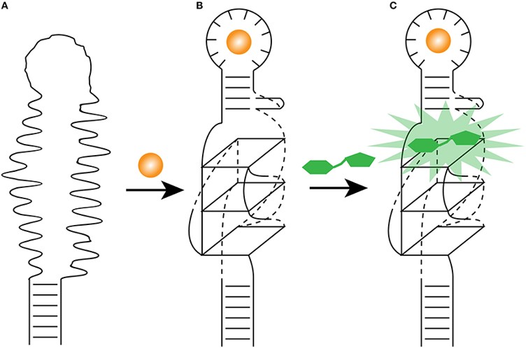 Fig.1 Light-up aptamer as biosensors.1,3
What Can We Offer?
We offer a comprehensive suite of development services designed to create high-performance fluorescent aptasensors. Our team handles everything from initial fluorophore selection and site-specific labeling to the engineering of advanced energy transfer systems for ultra-sensitive detection.
Custom Fluorophore & Quencher Integration
We provide seamless integration of standard organic dyes and dark quenchers during solid-phase synthesis. Our expertise ensures that labels are placed at positions that maximize structural stability and signal recovery.
One-Pot Aptamer-Quantum Dot Synthesis
We offer a specialized hydrothermal route where the aptamer serves as the stabilizing ligand during Quantum Dot growth. This results in ultra-small (<10 nm), bio-stable probes with high quantum yields and reduced cytotoxicity compared to traditional conjugation methods.
Site-Specific Post-Synthetic Labeling
For aptamers requiring modifications not compatible with automated synthesis, we employ click chemistry (Azide-Alkyne) or thiol-maleimide coupling to achieve precise, 1:1 labeling ratios.
Conformational Switch Optimization
Our scientists optimize the sequence to ensure a dramatic structural transition upon target binding, such as the transition from a random coil to a G-quadruplex, which is critical for "Turn-On" fluorescence applications.
Workflow
01
Technical Consultation: We discuss your target (protein, small molecule, or cell), required sensitivity, and detection environment (e.g., buffer vs. serum).
02
Aptamer Material Intake: Clients provide the aptamer sequence or the raw aptamer candidates from a SELEX pool.
03
Design & Simulation: We determine the optimal labeling sites (5', 3', or internal) using secondary structure prediction to minimize "Affinity Cost."
04
Synthesis & Modification: We perform the modification using either solid-phase synthesis or our proprietary "One-Pot" hydrothermal quantum dots conjugation.
05
Purification & Validation: Probes are purified via HPLC/PAGE. We validate the signal-to-noise ratio and Kd via fluorescence titration.
06
Final Delivery: You receive the purified, lyophilized fluorescent aptamer, accompanied by a comprehensive QC report including MS/HPLC traces and fluorescence recovery data.
Published Data
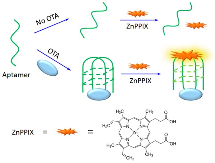 Fig.2 The design of OTA detection based on the structure switch of OTA aptamer.2,3
In this study, a label-free fluorescence sensing platform was developed for the highly sensitive detection of Ochratoxin A (OTA). The mechanism utilizes the OTA-induced conformational transition of a specific aptamer sequence into a G-quadruplex structure. In the presence of the fluorescent probe Zinc(II)-protoporphyrin IX (ZnPP), which specifically binds to G-quadruplexes, a significant enhancement in fluorescence signal is observed upon target recognition. The project achieved an exceptional detection limit of 0.03 nM, demonstrating high selectivity over other competitive mycotoxins. This research underscores the ability to engineer aptasensors that leverage structural switches for robust performance in complex food matrices, such as corn and red wine.
Why Choose Us?
-
Scientific Management: Our labs are managed by PhD-level scientists who provide collaborative, peer-to-peer interaction for every project.
-
Ultra-Small Probe Dimensions: Our "One-Pot" synthesis produces probes significantly smaller than antibody-based reporters, facilitating superior cellular uptake.
-
Affinity Preservation: We specialize in minimizing the "Affinity Cost," ensuring that fluorescence modifications do not interfere with the aptamer's binding equilibrium.
-
Cost-Efficient Production: We utilize optimized synthesis protocols to maintain the highest quality standards while reducing the time and cost of production.
Applications
-
Real-Time Biosensing: Development of ultrasensitive "Turn-On" sensors for the dynamic detection of metabolites, small molecules, and disease-related proteins in raw biological fluids without extensive sample preparation.
-
High-Throughput Screening (HTS): Fluorescently modified aptamers serve as highly effective displacement probes for screening massive small molecule libraries, accelerating drug discovery by identifying potential inhibitors through rapid, cost-effective optical readouts.
-
Intracellular Imaging and Tracking: Utilizing photostable Quantum Dot-Aptamer conjugates to track protein expression, organelle localization, and molecular trafficking within live cells with high spatial resolution and minimal photobleaching.
-
Food Safety and Environmental Monitoring: Rapid, often visual detection of regulated mycotoxins, heavy metal contaminants, and pathogens in complex matrices like grain extracts or environmental water samples, ideal for point-of-care testing.
FAQs
Q: How do you ensure the fluorophore doesn't inhibit the aptamer's binding affinity?
A: We utilize advanced secondary structure modeling to identify specific "loops" or termini that are not involved in target recognition. By placing the fluorophore in these non-essential regions, we minimize the "Affinity Cost." If binding is impacted, we can introduce a flexible PEG spacer between the aptamer and the dye to reduce steric hindrance.
Q: Can you perform internal modifications, or are labels restricted to the 5' and 3' ends?
A: We offer both options. While 5' and 3' labeling is most common and cost-effective for most researchers, we can incorporate fluorescently modified nucleotides (e.g., Fluorescein-dT) internally during synthesis. This is often necessary for specialized FRET-based molecular beacons where the distance between the donor and acceptor must be precisely controlled.
Q: Do you offer modifications that improve the stability of the fluorescent aptamer in serum?
A: Yes, we recommend a "Stability Package" alongside fluorescence modification. This includes 2'-Fluorine or 2'-O-Methyl ribose modifications and 3'-inverted thymidine capping. These structural changes prevent nuclease degradation, ensuring your fluorescent signal accurately reflects target binding rather than non-specific probe breakdown in biological matrices.
Q: What is "One-Pot" synthesis, and why should I choose it over standard EDC/NHS coupling?
A: In One-Pot synthesis, the aptamer is present during the actual formation of the Quantum Dot. This creates a direct, robust bond and ensures that the aptamer is perfectly oriented on the surface. Traditional coupling can lead to random orientation and larger hydrodynamic sizes, which can significantly interfere with cell penetration and binding kinetics.
Q: Can I use your fluorescent aptamers for multiplexed detection?
A: Absolutely. We can synthesize a custom panel of aptamers labeled with dyes that have non-overlapping emission spectra (e.g., FAM, Cy3, and Cy5). This allows you to monitor multiple unique targets simultaneously within a single sample or cell without signal crosstalk.
Creative Biolabs' expertise in fluorescence modification empowers your research with ultra-sensitive, real-time molecular recognition tools. Our scientists are ready to help you design the perfect optical probe for your specific application. Contact us today to discuss your project requirements.
Featured Services
Featured Products
References
-
Ouellet, Jonathan. "RNA fluorescence with light-up aptamers." Frontiers in Chemistry 4 (2016): 29. https://doi.org/10.3389/fchem.2016.00029
-
Liu, Feng, et al. "A label-free aptasensor for ochratoxin A detection based on the structure switch of aptamer." Sensors 18.6 (2018): 1769. https://doi.org/10.3390/s18061769
-
Distributed under Open Access license CC BY 4.0, without modification.
For Research Use Only.
Related Sections: