HSC activation relies on Wnt/β-catenin signaling, driving proliferation and profibrotic mediator secretion like plasminogen activator inhibitor-1 (PAI-1). PAI-1 inhibits ECM degradation. Our assays quantify your compound's inhibition of β-catenin nuclear translocation and suppression of PAI-1 expression (mRNA/protein), linking efficacy to restored ECM turnover.
LX-2 Cell based Antagonist Assay Service
At Creative Biolabs, we have leveraged the immortal LX-2 cell line, a human hepatic stellate cell (HSC) line, to develop advanced antagonist assays aimed at evaluating potential antifibrotic compounds. LX-2 cells preserve the functional characteristics of in vivo HSCs and are used to screen inhibitors effectively through high-content analyses. These assays provide precise quantifications of the fibrogenic responses and antagonistic effects of test compounds, solidifying our position at the forefront of fibrosis research.
Background What We Can Offer Workflow Publication Why Choose Us FAQs Customer Review Related Services Contact Us
Introduction: LX-2 Cell-based Antagonist Assay Fundamentals
Liver fibrosis is fundamentally defined by the activation of quiescent HSCs into highly contractile, matrix-producing myofibroblasts. This transdifferentiation process leads to the pathogenic, excessive deposition of extracellular matrix (ECM), primarily collagen type I. Our service utilizes the immortalized human LX-2 cell line as the industry standard model for HSC activation due to its human origin, reliable proliferative capacity, and retention of critical cytokine receptors. The assay is strategically configured to interrogate multiple recently validated profibrotic pathways, including Wnt/PAI-1, RNF2/MAPK (ERK/p38), and cytokine-mediated retinoid dysregulation (LRAT/CRBP1), providing high-fidelity, human-relevant data for candidate validation.
Key Deliverables Through Mechanistic Targeting
Targeting the Wnt/β-catenin and PAI-1 Axis
Analyzing the RNF2/MAPK Signaling Cascade
The E3 ubiquitin ligase RNF2 promotes fibrogenesis by activating the MAPK pathway. We measure phosphorylation inhibition of downstream kinases ERK1/2 and p38, providing mechanistic validation of your candidate as an upstream signal modulator.
Validating Cytokine-Mediated Retinoid Dysregulation
Activated HSCs lose vitamin A stores, driven by cytokines like TNF-α and IL-1β that suppress key enzymes LRAT and CRBP1. Our service assesses your compound's ability to preserve these markers, offering early validation of HSC deactivation potential.
Ready to accelerate your antifibrotic pipeline? Contact us today to discuss how our high-content LX-2 assay can validate your lead compounds and move your program closer to the clinic.
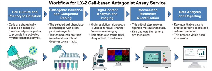
Workflow for LX-2 Antagonist Screening
Our assay is meticulously designed to provide robust and reproducible results, allowing for the reliable quantification of a test compound's ability to prevent or reverse the fibrogenic process.
Publication
This study investigates the anti-fibrotic mechanism of eupatilin, a natural flavonoid, in hepatic fibrosis. Using the human hepatic stellate cell line LX-2 and a CCl4-induced mouse model, the research demonstrates that eupatilin significantly alleviates liver fibrosis. It suppresses HSC activation, proliferation, and the epithelial-mesenchymal transition (EMT) process. The underlying mechanism involves the inhibition of the β-catenin pathway, reducing its nuclear translocation and subsequently downregulating its key downstream target, plasminogen activator inhibitor-1 (PAI-1). This work identifies eupatilin as a promising therapeutic agent and a natural PAI-1 inhibitor for treating hepatic fibrosis.
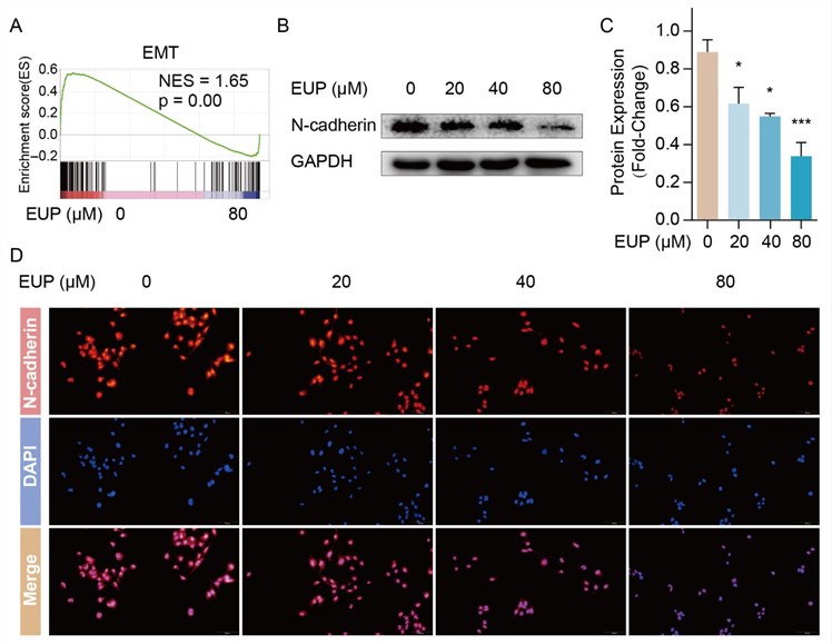 Fig. 1 Eupatilin suppresses epithelial-mesenchymal transition in LX-2 cells. 1
Why Choose Us
Creative Biolabs is your specialized partner in antifibrotic drug discovery, offering unparalleled depth in liver fibrosis modeling. Our advantage lies in combining the human-relevant LX-2 cell model with advanced, multi-mechanistic readout capabilities targeting underlying drivers of HSC activation. We offer Deep Mechanistic Validation targeting three critical pathways and customizable phenotype selection. Utilizing cutting-edge imaging platforms for detailed, multiparametric data, our integrated service offering bridges LX-2 assays with follow-up 3D spheroid models. We provide validated, actionable data that significantly accelerates your drug development decisions.
Experience the Creative Biolabs Advantage - Get a Quote Today
FAQs
Can your LX-2 assay distinguish between a compound that kills activated HSCs and one that genuinely reverses the fibrotic phenotype?
Absolutely. Our service incorporates multiplexed analysis, including a dedicated viability assay, alongside tracking specific deactivation markers and proliferation markers. This multiparametric approach ensures you confidently distinguish true antifibrotic action from non-specific cytotoxicity.
What is the main advantage of using your LX-2 model compared to primary human or rodent HSCs for high-throughput screening?
While primary cells offer undeniable biological relevance, LX-2 cells provide superior reproducibility, high availability, and a standardized human genetic background necessary for HTS and reliable IC50 determination.
How do you confirm the activation of the three different pathways in the LX-2 cells simultaneously?
TGF-β1 drives the classical fibrotic response, while the inclusion of inflammatory cytokines like TNF-α is necessary to fully trigger the retinoid dysregulation pathway. We rigorously confirm activation by validating the expected shifts in key markers before any compound screening begins.
Customer Review
-
Mechanistic Clarity
The multiplexed readout, particularly the p-ERK/p38 data, helped us confidently distinguish our potent hit from weaker candidates that only showed generalized toxicity, accelerating our decision-making. - Dr. Al*n W -
Quiescent Model Utility
The matrigel-seeded quiescent model accurately replicates the native HSC phenotype, giving us confidence that our hit compound is truly preventing activation, not just killing cells. - Dr. El*a K
Related Services
To ensure a seamless transition from lead identification to preclinical validation, Creative Biolabs offers several complementary services that can be integrated with your LX-2 antagonist assay:
Fibrosis Biomarker Panel Screening Service
Our service provides in-depth fibrosis biomarker analysis via high-throughput assays and cellular models, accelerating the discovery of novel targets and the evaluation of therapeutic candidates.
Learn More →Drug Metabolic Stability Analysis Service
Creative Biolabs offers in vitro metabolic stability assays to predict drug behavior in vivo. This service identifies therapeutic drugs with optimal metabolic stability, ensuring favorable bioavailability and reduced toxicity risk.
Learn More →Contact Us
Creative Biolabs is dedicated to providing specialized, data-driven solutions that accelerate the development of groundbreaking antifibrotic therapies. Our LX-2 cell-based antagonist assay service provides the precision and depth required to make critical decisions about your pipeline.
Contact Our Team for More Information and to Discuss Your Project
Reference
- Hu, Jinyuan, et al. "Eupatilin ameliorates hepatic fibrosis and hepatic stellate cell activation by suppressing β-catenin/PAI-1 pathway." International Journal of Molecular Sciences 24.6 (2023): 5933. Distributed under Open Access license CC BY 4.0, without modification. https://doi.org/10.3390/ijms24065933
 Download our brochure
Loading case studies...
Online Inquiry