Introduction of CR2
Complement receptors are expressed either on or in the cells of the innate and the adaptive immune systems. Complement receptors control the recruitment of blood leucocytes to the sites of inflammation, promote phagocytosis, and/or extracellular killing of microorganisms by immune cells. Moreover, they are used for the clearance of immune complexes generated during infectious or non-infectious inflammatory events. Furthermore, complement receptor activation controls the development of adaptive immune responses towards pathogens, allergens, autoantigens, and altered self-molecules.
Complement receptor type 2 (CR2 or CD21), part of the B-cell receptor (BCR) co-receptor complex, binds iC3b and C3dg and further promotes differentiation of activated B cells to antibody-secreting plasma cells; thus, CR2 is the principal complement receptor that regulates B-cell responses and enhances B-cell immunity. CR2 binds to downstream activation products of C3, and is reactive with iC3b, C3dg with higher affinity than with C3d, but not bind to C3b.
Structure of CR2
CR2 is a type I single-chain transmembrane glycoprotein of size in 140-145 kDa. CR2 has an extracellular domain composed entirely of 15-16 SCRs (depend on allotypes), a 24 amino acid transmembrane region, and a 43 amino acid cytoplasmic tail. SCR2 and SCR1 of the extracellular domain constitute the primary site interacting with C3d, and the SCR3 and SCR4 exhibit low affinity for C3b and inactive form of C3. Besides interaction with complement, CR2 also associates with IFN-α, the low-affinity IgE receptor CD23, deoxyribonucleic acid (DNA), and GPs gp350and gp220 of EBV. On B cells, CR2 can associate with CD19 and CD81 to form the co-receptor of the antigen-specific BCR.
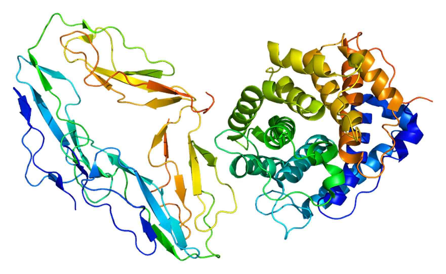
Fig. 1 CR2 3D structure.1
Cellular Distribution of CR2
Although CR2 is predominantly expressed by B lymphocytes, it can occur on other cell types. CR2 is strongly expressed on mature B lymphocyte cells (approximately 8000 CR2 molecules per cell) and lymph node follicular dendritic cells (FDCs), but absent on pre-, immature- or terminally differentiated plasma B cells. CR2 is to a lesser degree on activated T cells, K Cells with T cell markers lymphoblastoid cells, astrocytes, monocytes, neutrophils, basophils, thymocytes, and pharyngeal epithelial cells.
Biological and Signaling Functions
The intracellular cytoplasmic tail of CR2 itself is short and lacks apparent motifs to mediate direct signaling. However, as part of the BCR co-receptor complex, CR2 can associate with the B-cell surface molecule CD19 to signal with co-activator function. Thus, it ensures the recruitment of the latter to the BCR complex upon BCR/CR2 cross-linking by antigen engagement. Cross-linking CD19 to the BCR supplements the signaling transduced and lowers the B cell activation threshold by several orders of magnitude.
CR2 has been found to regulate both the effector cell function and the regulation of lymphocyte activation. There are a variety of products based on the CR2 which are offered by Creative Biolabs, including but not limited to: anti-CD21 monoclonal/ polyclonal antibodies, CR2 ELISA kits, CR2 immunizing/blocking peptides, CR2 protein lysate. CR2 on B cells participates in the phagocytose exogenously and the transport of antigens into secondary lymphoid organs, that is, from the subcapsular sinus into the lymph node or from the marginal zone into the spleen. CR2-based constructs, targeting C3d and delivering local complement regulatory activity, have been tested in diverse settings of tissue damage upon ischemia/reperfusion injury, chronic inflammation, and autoimmunity.
If you are interested in more information about complement receptor CR2, please feel free to contact us.
Reference
-
From Wikipedia: By Emw - Own work, CC BY-SA 3.0 https://commons.wikimedia.org/wiki/File:Protein_CR2_PDB_1ghq.png
Related Product
For Research Use Only.
Related Sections: