CR1 is a type I transmembrane glycoprotein of 2039 amino acids; the leader sequence is between amino acids 1 and 41. The mature peptide is located between position 42 and 2039, with a 25 amino acid transmembrane domain, between position 1972-1996, and with a 43 amino acid C-terminal domain, containing two PDZ motifs. There are four major structural allotypes recognized in humans: 1 (F, fast or A), 2 (S, slow or B), 3 (Cor F) and 4 (D).
The relative molecular mass of CR1 depends on the cellular source of CR1, type of electrophoresis system and buffers used. CR1 obtained from nucleated blood cells run higher than CR1 from red cells, due to altered N-glycosylation. Glycosylation contributes with 20-25 kDa to the molecular mass of CR1. There are 21 glycosylation sites on mature CR1 molecules.
Table1 The physicochemical properties of the mature protein
|
Allotype
|
Relative Molecular Mass
(Mr, kDa, Reduced)
|
Relative Molecular Mass
(Mr, kDa, Unreduced)
|
|
CR1*1
|
220-250
|
190-210
|
|
CR1*2
|
250-280
|
220-250
|
|
CR1*3
|
190-220
|
160-190
|
|
CR1*4
|
Over 280
|
Over 250
|
-
CR1 Structure
The extracellular domain of CR1 is comprised of 30 short consensus repeats (SCRs), SCRs are also known as complement control protein repeats or Sushi domains. Extracellular SCRs are further grouped in four, tandem large homologous repeats, LHR-A, LHR-B, LHR-C and LHR-D, each 45 kDa in size, and composed of seven SCRs. There are two unassigned SCRs near the plasma membrane, SCR 29 and 30. The homology between the four LHRs varies between 60% and 99%. The S variant of CR1, CR1*2, has an additional LHR-S inserted between LHR-B and LHR-C. The transmembrane region of CR1 consists of 25 hydrophobic amino acids followed by four positively charged residues and the C-terminal cytoplasmic domain consists of 39 amino acids. The transmembrane domain, higher primates, baboons, and macaques have GPI-anchored version of CR1 with identical extracellular portions.
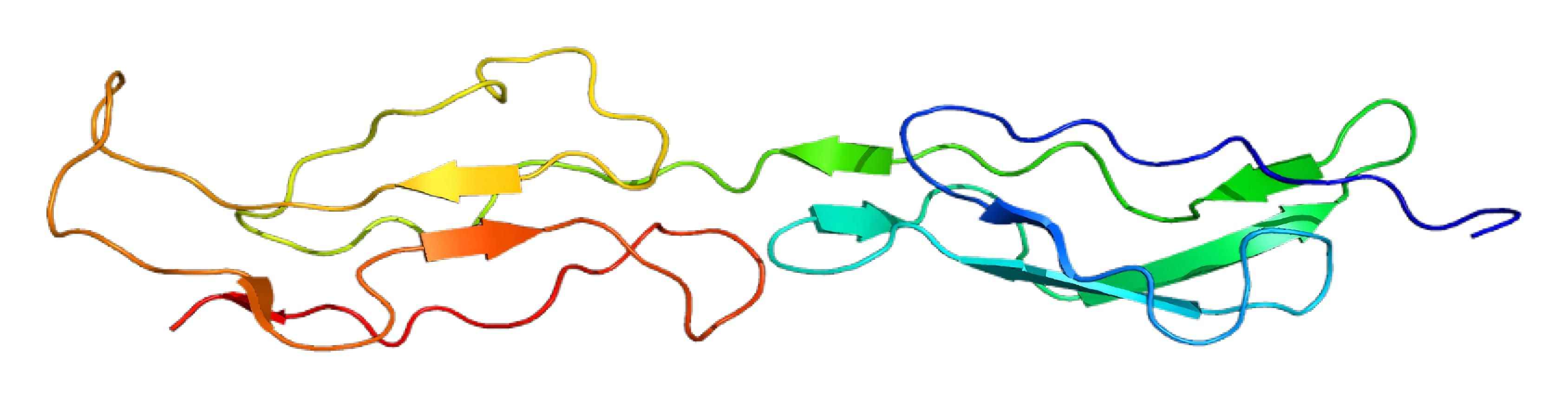
Fig. 1 Diagram of CR1 structure.1
-
CR1 Function
In humans, the main function of CR1 is binding of complement fragments complement C3b, complement factors C4b, C1q and MBL (mannan-binding protein). The interaction between CR1 and iC3b is weak. CR1 on circulating red blood cells (RBCs) binds complement-opsonized immune complexes and microbes present in blood and delivers them to tissue-resident macrophages in liver and spleen through a process called immune adhesion clearance. By preventing immune complexes to persist in circulation, and directly interact with nucleated blood cells, RBC CR1 contributes to maintaining an anti-inflammatory environment in blood.
On T cells and dendritic cells, CR1 participates in antigen presentation. In RBCs, CR1 promotes calcium ion influx and increases membrane deformability. CR1 modulates B cell function and is involved in the polarisation of Tregs. The complement regulatory function of CR1 depends on soluble factor I, which acts as a cofactor during degradation of complement fragment C3b to iC3b and C3f, and of iC3b to C3c and C3d. In the presence of factor I, CR1 also cleaves C4b to C4c and C4d and accelerates the degradation of the C3 and C5 convertases of the classical and alternative complement activation pathways.
CR1 Tissue Distribution
In blood, CR1 is expressed on RBCs, neutrophils, monocytes, B cells, subpopulation of T and activated NK cells. In tissue, CR1 is found on podocytes, activated endothelial cells, neurons, astrocytes, and follicular dendritic cells. A soluble form of CR1, with concentration ranging 13-81 ng/mL, exists in plasma that could be either actively secreted by the cells, likely circulating neutrophils, or the product of enzymatic cleavage. The soluble form of CR1 may play a protective role during inflammatory conditions.
Creative Biolabs is committed to providing high-quality complement component-related services and products. If you are interested in our services, please directly contact us and consult our technical supports for more details.
Reference
-
From Wikipedia: By Emw - Own work, CC BY-SA 3.0 https://commons.wikimedia.org/wiki/File:Protein_CR1_PDB_1gkg.png
Related Product
For Research Use Only.
Related Sections: