iPSC Retinal Pigment Epithelium Ex Vivo Modeling Service
Accelerate Your Retinal Research with Unparalleled Precision!
Are you currently facing challenges in accurately modeling retinal diseases, developing effective retinal therapies, or understanding the complex functions of the retinal pigment epithelium (RPE)? Our iPSC Retinal Pigment Epithelium Ex Vivo Modeling Service helps you accelerate your research and obtain physiologically relevant models through advanced iPSC differentiation and characterization.
How Creative Biolabs' iPSC Retinal Pigment Epithelium Ex Vivo Modeling Service Can Assist Your Project?
Creative Biolabs' iPSC Retinal Pigment Epithelium Ex Vivo Modeling Service provides you with a robust and reliable platform for studying retinal biology, disease mechanisms, and therapeutic interventions. We deliver high-quality, functional RPE models tailored to your specific research needs.
Workflow:
Our streamlined workflow ensures efficient and reliable generation of iPSC-derived RPE models:
Project Consultation and Planning
We begin with a detailed consultation to understand your research objectives, design, and desired outcomes, including the specific disease model, assays, and characterization methods required. Clients typically provide patient-derived or other relevant iPSC lines, disease information, and functional assay requirements.
iPSC Culture and Expansion
We employ advanced cell culture techniques to expand and maintain your iPSC lines, ensuring sufficient high-quality starting material. This step can also involve rigorous quality control to confirm iPSC pluripotency and viability (if needed).
RPE Differentiation
Our optimized protocols guide iPSCs through controlled steps to induce differentiation into RPE cells, mimicking the natural developmental pathway to ensure appropriate cell characteristics, backed by the excellent NeuroST™ platform.
Characterization and Quality Control
We perform a comprehensive suite of assays to validate RPE cell identity, purity, and functionality. This includes morphological assessment, immunofluorescence staining for RPE-specific markers, secretion of growth factors, and functional assays.
Data Analysis and Reporting
We analyze data from characterization assays and compile a detailed report summarizing results. Clients receive a comprehensive written report, high-resolution images, microscopy data, and quantified data from functional assays.
Delivery and Support
We deliver differentiated and characterized RPE cells, along with protocols for further culture and downstream applications, and provide ongoing technical support. The typical timeframe is 8 to 16 weeks, depending on project complexity, iPSC lines, and the extent of characterization.
Discover How We Can Help - Request a Consultation
Why Choose Us?
Creative Biolabs is a trusted provider of advanced cell-based solutions, with a proven track record of delivering high-quality results. Our iPSC Retinal Pigment Epithelium Ex vivo Modeling Service offers several key advantages:
Expertise
Our team comprises experienced scientists with deep expertise in iPSC technology, retinal biology, and cell differentiation. We deliver high-quality, functional RPE models tailored to your specific research needs. We can also generate physiologically relevant RPE models from patient-derived iPSCs that accurately reflect the genetic and phenotypic characteristics of the disease, enabling the specific disease research.
Customization
We tailor our services to meet your specific research needs, ensuring that you receive the precise RPE models and data you require. We offer a range of customizable assays, including phagocytosis assays and gene expression analysis, to evaluate RPE function.
Quality
We adhere to rigorous quality control standards at every step of the process, ensuring the reliability and reproducibility of our results. We deliver detailed reports with comprehensive data analysis, providing you with actionable insights into your research questions.
Advanced Technology
We utilize state-of-the-art equipment and optimized protocols to generate highly functional and physiologically relevant RPE models, powered by the cutting-edge NeuroST™ platform for neuroscience drug discovery.
Comprehensive Solutions
Creative Biolabs offers a wide range of complementary services, including iPSC generation, gene editing, and other cell differentiation services, providing you with a complete solution for your research needs. iPSC-derived RPE cells and retinal organoids can be used for a wide range of applications, including disease modeling, drug discovery, toxicology testing, and cell therapy development.
Our clients experience an average 90% success rate in generating functional RPE models and a 75% reduction in project timelines.
Case Study:
Creative Biolabs establishes the in vitro RPE differentiation platform from human iPSC and confirms marker gene expression (MiTF and ZO1) of matured PRE by IF staining. FACS further proves the high differentiation efficiency (≥ 95%). ELISA detection detects the secretion of VEGF and PEDF.
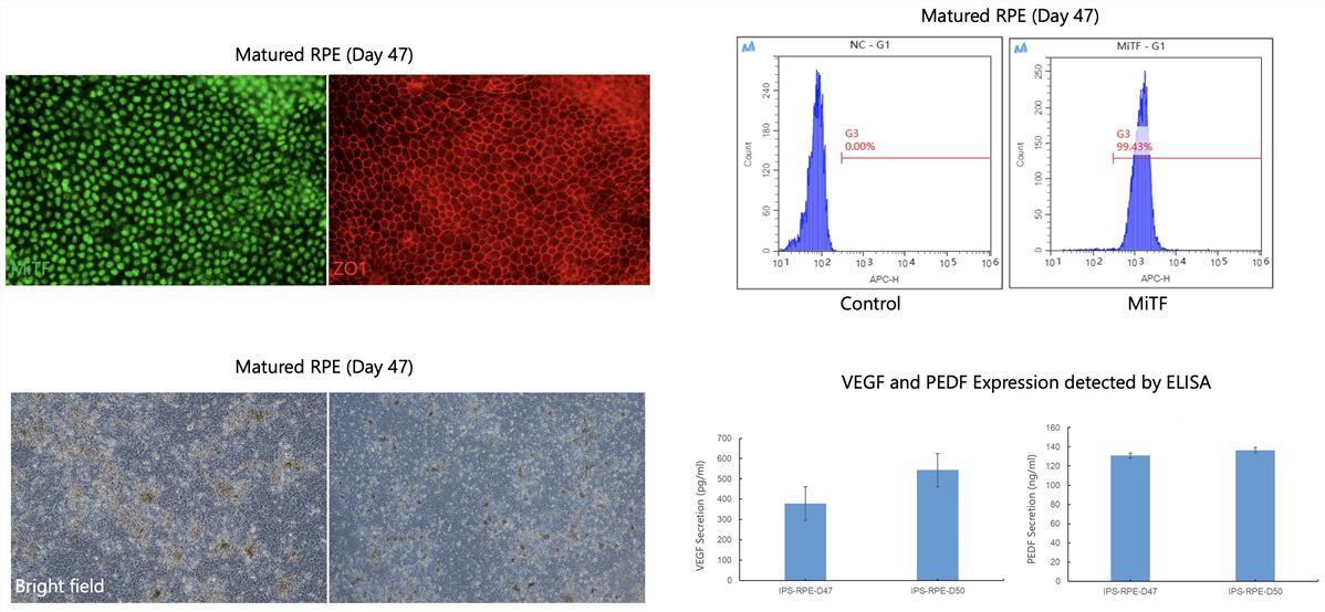 Fig.1 Human iPSC-derived matured RPE.
Customer Reviews:

Experience the Creative Biolabs Advantage - Get a Quote Today
Introduction
The retinal pigment epithelium (RPE) is a critical cell layer in the retina, playing a vital role in maintaining photoreceptor function and overall retinal health. Dysfunction of the RPE is implicated in a wide range of retinal diseases, including age-related macular degeneration (AMD) and inherited retinal degenerations. Recent advances in iPSCs technology have revolutionized the study of retinal diseases. iPSCs can be differentiated into RPE cells, providing a valuable in vitro model for investigating disease mechanisms, screening potential therapies, and developing new treatment strategies. The NeuroST™ platform from Creative Biolabs leverages these advancements.
FAQs
Here are some frequently asked questions from potential customers interested in our iPSC Retinal Pigment Epithelium Ex Vivo Modeling Service:
Q: Can you generate RPE models from iPSCs derived from any patient?
A: Yes, we can work with most patient-derived iPSC lines. However, we recommend that you contact us with the specific details of your iPSC lines, and our team will assess their suitability for RPE differentiation. This will allow us to give you the most accurate information and guidance for your project.
Q: What kind of functional assays can you perform on the RPE models?
A: We offer a range of functional assays, including phagocytosis assays, transepithelial electrical resistance (TEER) measurements, and cytokine secretion analysis. We can also customize assays to meet your specific research needs. Contact us to discuss your specific requirements.
Q: How do your iPSC-derived RPE models compare to traditional cell lines?
A: Our iPSC-derived RPE models offer several advantages over traditional cell lines, including a more physiologically relevant phenotype, better representation of disease-specific characteristics, and the ability to generate patient-specific models. These models, along with the NeuroST™ platform, provide more accurate and predictive results.
Q: What kind of data will I receive?
A: You will receive a comprehensive report that includes detailed information about the differentiation process, morphological characterization, biomarker expression analysis, and functional assay results. We provide high-quality images, quantitative data, and a clear interpretation of the findings.
Related Products
Creative Biolabs provides a readily accessible portfolio of high-quality differentiated cells derived from iPSCsp:
| Products | Donor | Size | Quality control |
|---|---|---|---|
| iPSC differentiated NPC cells | Healthy | 106 cells / vial |
≥ 80% PAX6+ cells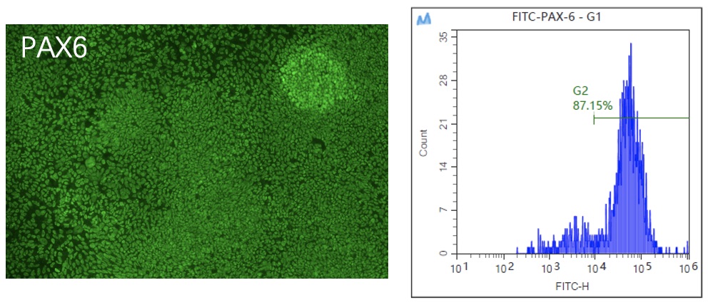
|
| iPSC differentiated RPE | Healthy | 106 cells / vial |
≥ 95% MiTF+ cells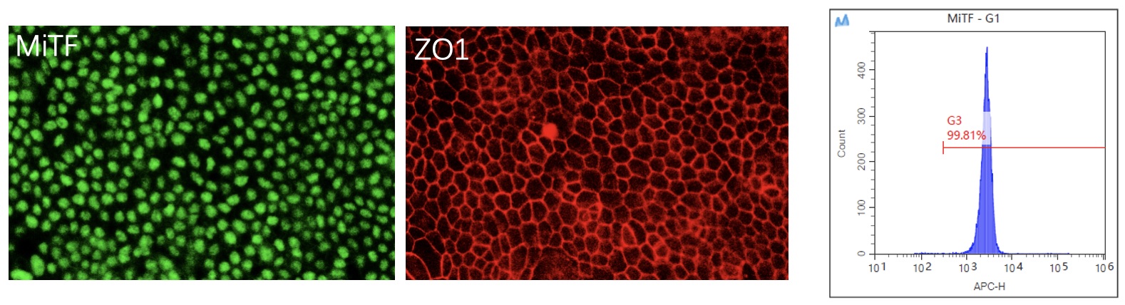
|
Related Services
Creative Biolabs offers an extensive array of solutions for neuroscience research. Beyond our iPSC Retinal Pigment Epithelium Ex Vivo Modeling Service, we also provide:
How to Contact Us
Creative Biolabs' iPSC Retinal Pigment Epithelium Ex Vivo Modeling Service provides a powerful and versatile tool for advancing your retinal research. Our expertise, customization options, and commitment to quality ensure that you receive the high-quality RPE models and data you need to accelerate your drug discovery, disease modeling, and cell therapy development efforts.
Contact our team today to learn more about how our iPSC Retinal Pigment Epithelium Ex Vivo Modeling Service and the NeuroST™ platform can support your research. We are ready to discuss your specific project requirements and provide you with a customized solution.
Contact Our Team for More Information and to Discuss Your Project