One-Stop
ASO Development
Services
From Sequence Design to Preclinical Support. We provide comprehensive solutions covering ASO design, synthesis, modification, conjugation, analysis, and in vivo/in vitro validation.
ASO Technology Overview
Synthetic Precision & Specificity
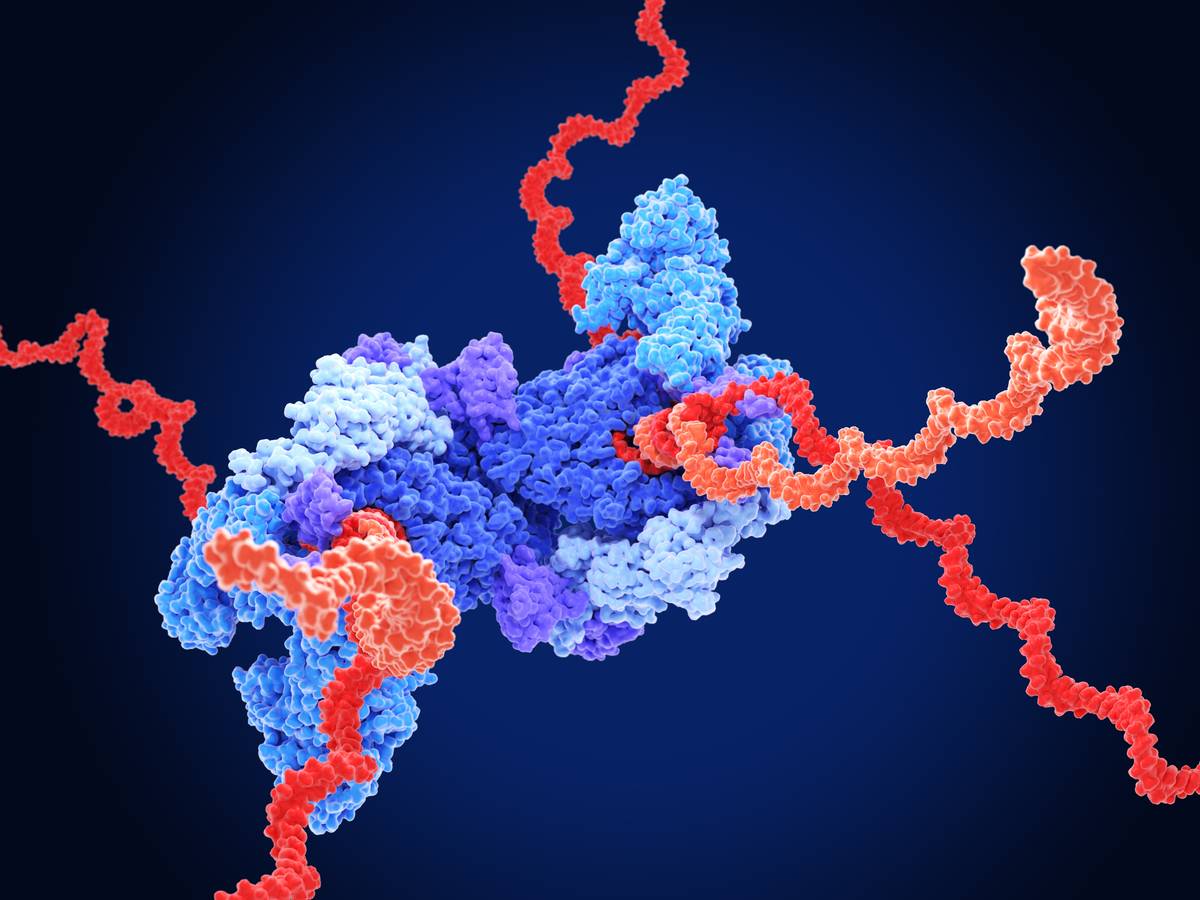
Antisense Oligonucleotides (ASOs) are short, synthetic, single-stranded oligodeoxynucleotides meticulously designed to alter RNA expression through precise base-pairing. By targeting the transcriptomic level rather than the proteome, ASOs provide a powerful therapeutic modality for addressing diseases that remain "undruggable" by conventional small molecules or biologics.
Our platform leverages advanced sequence optimization to ensure that each ASO candidate possesses maximal target affinity while maintaining a low off-target profile, enabling high-fidelity gene modulation.
Core Therapeutic Mechanisms
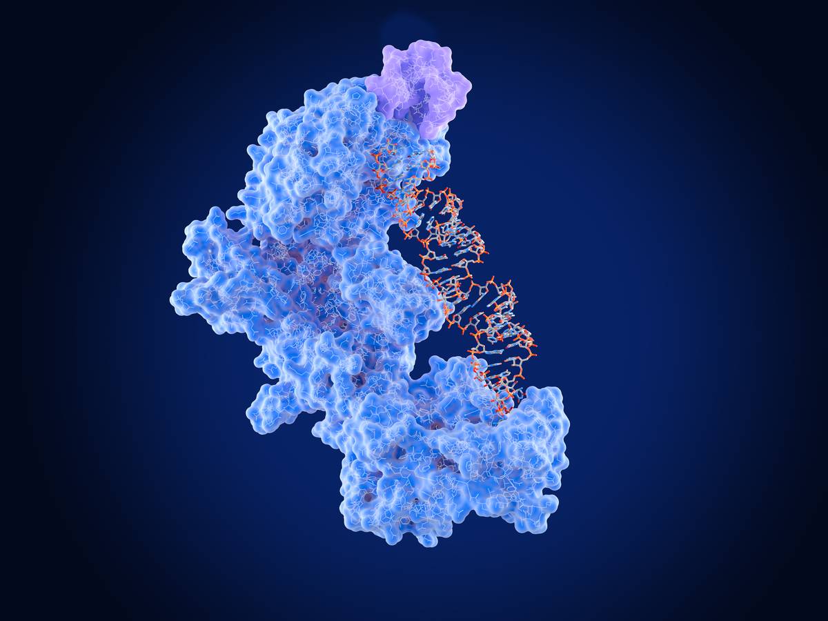
RNase H-Mediated Degradation
Primarily utilizing Gapmer designs to recruit RNase H1, leading to the cleavage and degradation of target mRNA for robust gene silencing. This approach employs a chimeric design with central DNA nucleotides flanked by chemically modified RNA analogues, which enables efficient enzyme recruitment while conferring nuclease resistance and enhanced target affinity.
Steric Blocking & Splice Modulation
Physically blocking translation or modulating alternative splicing (Exon skipping/inclusion) to restore functional protein expression or disrupt toxic RNA motifs. This strategy often uses fully modified oligonucleotides that avoid RNase H cleavage, instead exerting effects through precise hybridization to pre-mRNA or mRNA regions.
Genetic & Rare Disorders
Transformative potential for diseases such as SMA and DMD, where modulating RNA processing can fundamentally alter disease progression.
Oncology & Viral Infections
Targeting oncogenic drivers or viral transcripts that are otherwise inaccessible, providing a specialized toolset for precision medicine.
Neurodegenerative Diseases
Advanced ASO chemistry enables long-lasting effects in the CNS, tackling the root genetic causes of neurological decline.
One-Stop ASO Solutions
We offer a multi-dimensional development platform that integrates advanced bioinformatics, high-end chemical synthesis, and rigorous preclinical validation to ensure the success of your RNA therapeutic pipeline.
ASO Design & Sequence Optimization
Our design platform goes beyond basic base-pairing. We integrate RNA Secondary Structure Accessibility Prediction, using proprietary bioinformatic tools to identify "open regions" of the target RNA, thereby bypassing structural hindrance and ensuring high binding efficiency.
Thermodynamic Simulation (ΔG)
Predicting binding kinetics under physiological conditions to ensure superior affinity while minimizing homology-based off-target risks.
Structure-Based Design
Overcoming regional steric hindrance to unlock inaccessible RNA domains for higher knockdown potency.
Advanced Synthesis & Complex Modification
To maximize in vivo stability and reduce immunogenicity, we employ a sophisticated suite of chemical modifications. Our platform supports diverse architectures, including Gapmers for RNase H-mediated degradation and Steric-blocking oligos for splice modulation.
- Backbone: PS, PMO, Peptide-conjugated
- Sugar: 2'-OMe, 2'-MOE, cEt
- Scale: mg to multi-gram synthesis
- Ultra-high purity (HPLC/MS verified)
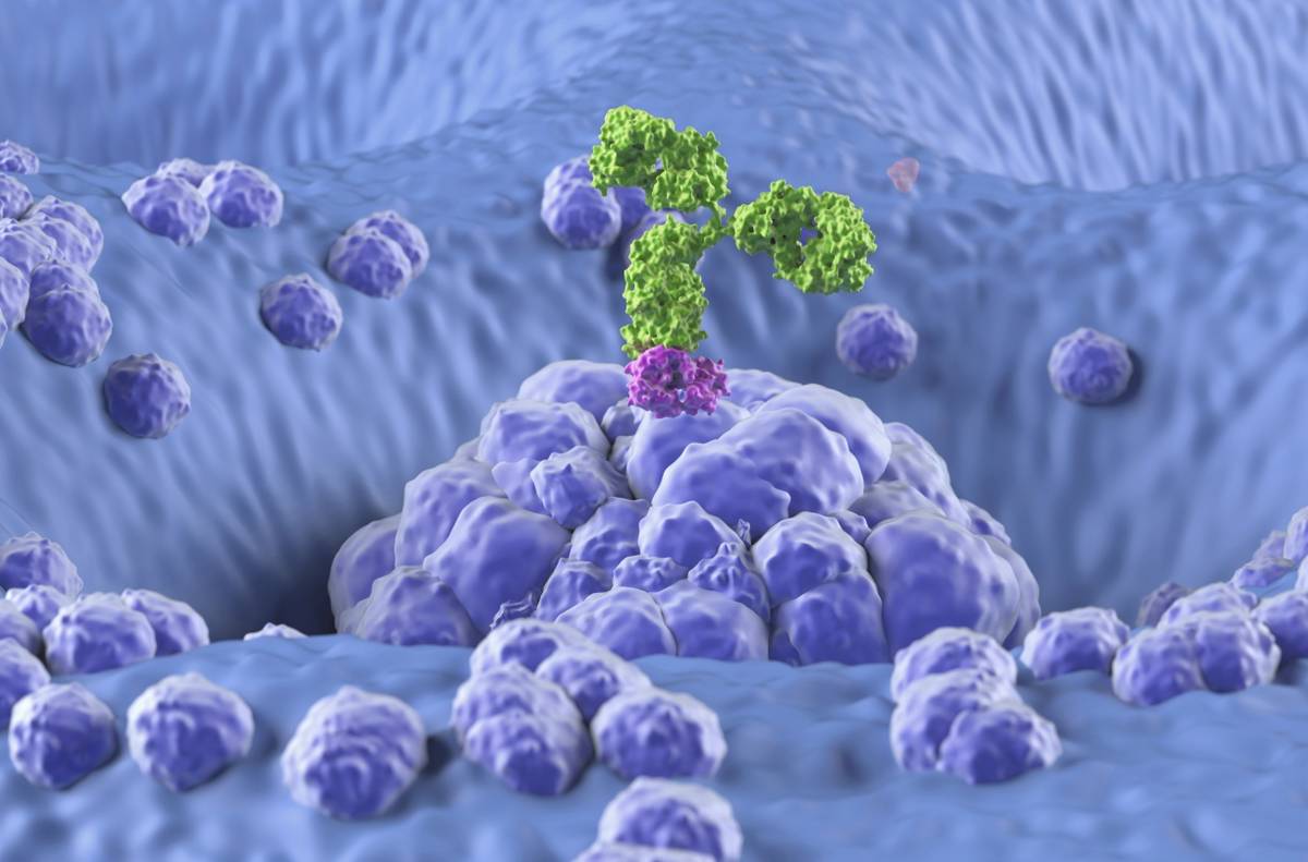
Precision Delivery & Conjugation
Overcoming the cellular uptake barrier is central to our strategy. We specialize in Targeted Conjugation technologies that enhance tissue specificity, significantly lowering the required dose and improving the safety profile of ASO therapeutics.
We match the conjugation chemistry to your specific indication, ensuring optimal PK/PD properties.
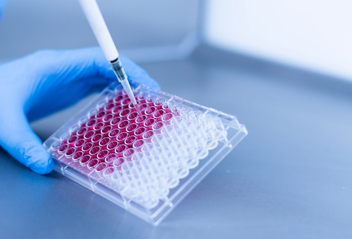
Integrated Preclinical Validation
Our validation pipeline provides a robust assessment of ASO performance. From high-throughput cell-based screening to in vivo proof-of-concept, we deliver high-quality data to support IND-enabling studies.
✓ In Vitro Efficacy
- • mRNA Knockdown (qPCR/bDNA)
- • Protein Level Analysis (WB/ELISA)
- • Cytotoxicity (MTT/CCK-8)
✓ In Vivo PK/PD
- • Tissue Distribution & Half-life
- • Biomarker Modulation
- • Tolerability & Safety Profiling
Who We Serve
Our flexible platform adapts to the needs of diverse partners, providing specialized technical support and strategic collaboration across the therapeutic development spectrum.
Biotech Companies
Strategic R&D Extension & Gap Bridging
We serve as a strategic R&D extension, providing end-to-end solutions that bridge the gap between initial hit discovery and preclinical proof-of-concept.
Allows lean teams to focus on core biology while we handle complex therapeutic development.
Pharmaceutical Enterprises
Modular Pipeline Support & Lifecycle Management
Offering modularized high-throughput screening and complex chemical modification platforms to support large-scale pipeline expansion.
Expertise in lifecycle management of established targets and advanced chemical synthesis.
Academic & Research
Translational Quality & Discovery Support
Translating fundamental genomic discoveries into therapeutic-grade candidates with rigorous QC standards.
Suitable for high-impact publications and meeting stringent translational grant requirements.
Startups (Incubation)
"Virtual Lab" Model for Early-Stage Innovation
We provide a "Virtual Lab" model allowing startups to execute sophisticated ASO R&D without building in-house infrastructure.
Eliminates prohibitive costs of internal synthesis and bioanalysis equipment.
Development Workflow
Our systematic workflow ensures high-quality data generation and technical continuity at every stage of your ASO drug discovery journey.
In Silico Design & Target Analysis
We begin with comprehensive bioinformatics modeling. This includes RNA secondary structure accessibility mapping and thermodynamic simulation (ΔG) to select the most potent and specific antisense sequences while screening for off-target risks.
Custom Synthesis & Modification
Custom synthesis of optimized leads using high-end phosphoramidite chemistry. We apply diverse chemical modifications 2'-MOE, LNA, PS backbone) to enhance metabolic stability and binding affinity, tailored to the specific target mechanism (RNase H or Steric Block).
Advanced Delivery
Conjugation
Implementation of targeted delivery strategies. This involves conjugating ASOs with GalNAc, Antibodies (AOCs), or cell-penetrating peptides to ensure tissue-specific uptake and efficient endosomal escape, optimizing the drug's PK/PD profile.
Cell-Based Efficacy
Screening
High-throughput validation in relevant cell models. We quantify mRNA knockdown efficiency via qPCR/bDNA and evaluate protein expression levels. Safety is assessed through comprehensive cytotoxicity and immunogenicity assays.
In Vivo
Proof-of-Concept
Transitioning to animal models to validate therapeutic efficacy. Studies include tissue distribution, biodistribution (ADME), dose-response relationships, and preliminary safety/tolerability assessments in living systems.
Bioanalysis & Preclinical Reporting
Comprehensive bioanalytical support including LC-MS/HPLC purity analysis and stability testing. We provide a full technical data package and methodology summary to support your studies and regulatory filings.
Bioanalysis & QC Support
Rigorous analytical characterization and stability testing are the cornerstones of successful ASO development, ensuring therapeutic integrity and regulatory compliance.
Analytical Characterization
-
●
Purity & Integrity Analysis
High-resolution LC-MS and HPLC-based purity verification (Ion-pair RP-HPLC / AEX-HPLC) to ensure consistent ASO quality.
-
●
Stability in Biological Matrices
Exonuclease and endonuclease resistance assays in simulated serum, plasma, and lysosomal environments (37°C incubation).
-
●
Quantitative Bioanalysis
Development of sensitive methods (e.g., hybridization-based ELISA or LC-MS/MS) for ASO quantification in tissues and biofluids.
Project Delivery
-
●
IND-Enabling Data Packages
Comprehensive documentation of lead optimization, including structural confirmation, sequence fidelity, and binding kinetics.
-
●
Full Technical Reports
Detailed methodology summaries and raw data integration to facilitate seamless regulatory filings and audit trails.
-
●
Methodology Validation
Standardized protocols for off-target assessment and immunogenicity profiling, ensuring data reliability for translation.
Why Partner with Us?
One-Stop Platform
Seamless integration from bioinformatics design and chemical synthesis to in vivo proof-of-concept.
Advanced Chemistry
Rich experience in complex modifications (2'-MOE, LNA) and site-specific conjugation technologies.
Tailored Strategies
Bespoke ASO design and delivery solutions optimized for your specific target and therapeutic indication.
Rigorous Quality
Comprehensive QC systems including LC-MS and HPLC to ensure maximum purity and sequence fidelity.
Targeted Delivery
Specialized platforms for GalNAc, AOC, and ARC to ensure tissue-specific uptake and safety.
Bioinformatics Design
Proprietary algorithms for RNA structure accessibility and off-target risk assessment.
IND-Enabling Data
Robust preclinical data packages covering qPCR, Western Blot, and PK/PD profiling.
IP Confidentiality
Strict adherence to confidentiality and IP ownership protocols to protect your innovation.
Frequently Asked Questions
Start Your Project Today
Tell us about your project, and our experts will get back to you with a customized quote and proposal.