Immune Cell Immunophenotyping Services
Immune Cell Immunophenotyping
Immunophenotyping is the precise identification and quantification of immune cell populations based on the expression of specific protein markers or unique molecular signatures. This high-resolution analysis of the immune landscape is indispensable across the full spectrum of biomedical research. In immuno-oncology, for instance, defining the composition of tumor-infiltrating lymphocytes (TILs)—such as the ratio of cytotoxic CD8+ T cells to immunosuppressive regulatory T cells (Tregs)—is a critical determinant of patient prognosis and response to checkpoint inhibitor therapies. In autoimmune diseases like lupus or multiple sclerosis, tracking shifts in B cell and T cell subsets provides essential pharmacodynamic data to assess the efficacy of novel immune-modulating agents. At Creative Biolabs, we leverage our advanced platforms to provide this foundational data, enabling a granular understanding of the mechanism of action for new therapeutics and the identification of robust biomarkers for patient stratification.
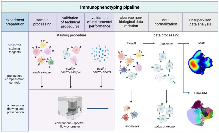 Fig.1 Immunophenotyping using high-dimensional flow cytometry and unsupervised data analysis.1
Our Comprehensive Immune Cell Immunophenotyping Service Content
Recognizing that no single technology can capture the full complexity of the immune system, Creative Biolabs offers an integrated suite of state-of-the-art platforms and targeted services. This multi-modal approach ensures that the optimal technology is applied to answer each specific scientific question, from broad surveillance to deep dives into specific cell lineages.
Targeted Immunophenotyping Services for Key Cell Lineages:
T Lymphocyte (T Cell) Immunophenotyping Service
Comprehensive analysis of CD4+ helper, CD8+ cytotoxic, and regulatory T cell (Treg) subsets. We provide deep profiling of memory and effector populations (Naive, Tcm, Tem, Temra), activation status, and exhaustion phenotypes through key markers like PD-1, CTLA-4, TIM-3, and LAG-3.
B Lymphocyte (B Cell) & Plasma Cell Immunophenotyping Service
Detailed characterization of the humoral immune response, including naive, memory (switched and unswitched), and transitional B cells. We quantify antibody-secreting cells, from plasmablasts to terminally differentiated, long-lived plasma cells (CD19, CD20, CD27, CD38, CD138).
Natural Killer (NK) Cell Immunophenotyping Service
Profiling of cytotoxic (CD56dim) and immunoregulatory (CD56bright) NK cell subsets. Our panels assess the expression of key activating and inhibitory receptors (e.g., NKG2D, KIRs) to determine the functional potential of the NK cell compartment.
Dendritic Cell (DC) Immunophenotyping Service
Precise identification of the primary antigen-presenting cell populations, including conventional DCs (cDC1 and cDC2) and plasmacytoid DCs (pDCs), which are critical for initiating and shaping adaptive immune responses.
Monocyte Immunophenotyping Service
Classification of circulating monocyte subsets—classical (CD14++CD16-), intermediate (CD14++CD16+), and non-classical (CD14+CD16++)—to understand their roles in inflammation, phagocytosis, and tissue repair.
Macrophage Immunophenotyping Service
Analysis of macrophage polarization states within tissues, distinguishing between pro-inflammatory (M1-like) and anti-inflammatory/pro-resolving (M2-like) phenotypes using markers such as CD80, CD86, CD163, and CD206.
Granulocyte Immunophenotyping Service
Quantification and characterization of neutrophils, eosinophils, and basophils. Our services can assess activation state, degranulation markers, and cellular purity for functional studies.
Myeloid-Derived Suppressor Cell (MDSC) Immunophenotyping Service
Critical for immuno-oncology research, this service differentiates between monocytic (M-MDSCs) and polymorphonuclear (PMN-MDSCs) subsets, quantifying their presence in the periphery and the tumor microenvironment.
Platelet Immunophenotyping Service
Analysis of platelet activation status via surface markers like P-selectin (CD62P) and their capacity to form aggregates with leukocytes, providing insight into thrombo-inflammation.
Red Blood Cell Immunophenotyping Service
Characterization of erythrocyte surface antigens for transfusion medicine and research, as well as markers of cellular senescence and health.
Mucosal Immune System Immunophenotyping Service
Specialized analysis of immune cells from gut, lung, or other mucosal biopsies, including tissue-resident memory T cells (TRM), intraepithelial lymphocytes (IELs), and lamina propria lymphocytes.
Fibrocyte Immunophenotyping Service
Identification of these unique bone marrow-derived mesenchymal progenitors that co-express hematopoietic (CD45) and stromal (collagen I) markers, which are implicated in fibrosis and wound healing.
Our Core Technology Platforms:
- High-Parameter Flow Cytometry: This remains the gold standard for high-throughput quantification of cell populations from blood and dissociated tissues. Our validated panels, using up to 20 distinct fluorescent markers, allow for the deep profiling of major immune lineages and their functional subsets.
- Single-Cell RNA Sequencing (scRNA-seq): Moving beyond protein expression, our scRNA-seq services profile the entire transcriptome of individual cells. This technology is transformative, allowing not only for definitive cell identification but also for the elucidation of a cell's functional state and developmental trajectory.
- Multiplex Immunohistochemistry (mIHC): Understanding cellular composition is often incomplete without spatial context. Our mIHC platform allows for the visualization of multiple immune cell types simultaneously within the native architecture of tissue sections, revealing the organization of immune "niches".
Our Advantages
Choosing Creative Biolabs provides access to a unique combination of technological innovation and deep scientific acumen, ensuring the highest quality data and interpretation.
- Advanced Epigenetic Analysis
- Integrated Computational Biology
- Unwavering Commitment to Quality
FAQs
Q1: Which technology is best suited for my study?
A1: The optimal technology depends entirely on your scientific question. For routine monitoring of major immune subsets in blood, flow cytometry is often sufficient. To discover novel cell populations or deeply characterize a response, scRNA-seq or CyTOF are superior. If the spatial location of cells within a tissue is critical, mIHC is the necessary choice. Our scientific consultants will work with you to design the most effective experimental plan.
Q2: What are the sample requirements for your services?
A2: We are highly flexible and can work with a wide range of sample types, including whole blood, PBMCs, dissociated tissue, fine-needle aspirates, and FFPE tissue blocks. For our epigenetic analysis platform, samples can be shipped ambient or frozen, significantly simplifying logistics.
Q3: How is the data delivered and analyzed?
A3: Clients receive a comprehensive data package including raw data files, processed quantification tables, and a detailed report from our bioinformatics team. The report includes high-resolution figures, statistical analysis, and a biological interpretation of the key findings, prepared in consultation with your team.
Contact Us
To advance your research with the highest level of precision in immune cell characterization, partner with Creative Biolabs. Our scientific team is ready to discuss your project-specific needs and develop a customized analytical strategy. Please reach out to our scientific liaison team to schedule an initial consultation and discover how our services can empower your next breakthrough.
Reference
- De Graaf, J Fréderique, and Ramon Arens. "Immunophenotyping beyond the limits of time." Cell reports methods vol. 3,10 (2023): 100612. DOI: 10.1016/j.crmeth.2023.100612. Distributed under Open Access License CC BY 4.0, without modification.
 Download our brochure