
Creative Biolabs is offering the most comprehensive services for antibody development projects. With strict regulation and effective execution, we are dedicated to providing the most valuable solutions to complete your projects.
HighlightsCreative Biolabs is offering the most comprehensive services for antibody development projects. With strict regulation and effective execution, we are dedicated to providing the most valuable solutions to complete your projects.
HighlightsCreative Biolabs has established a broad range of platforms for developing novel antibodies or equivalents. These cutting-edge technologies enable our scientists to meet your demands from different aspects and tailor the most appropriate solution that contributes to the success of your projects.
HighlightsWith deep understanding in antibody-related realms and extensive project experience, Creative Biolabs offers a variety of references to help you learn more about our capacities and achievements, including infographic, flyer, case study, peer-reviewed publications, and all kinds of knowledge that can assist your projects. You are also welcome to contact us directly for more specific solutions.
HighlightsGet a real taste of Creative Biolabs, one of the most professional custom service providers in the world. We are committed to providing highly customized comprehensive solutions with the best quality to advance your projects.
Highlights Loading...
All listed services and products are For Research Use Only. Do Not use in any diagnostic or therapeutic applications.
Platelet-derived growth factor subunit B(PDGFB) is a cysteine knot type growth factor subunit encoded by PDGFB gene, which is mainly synthesized and secreted by activated platelets, vascular endothelial cells and glioma cells, and released into extracellular microenvironment in the form of PDGF-BB homodimer. Compared with PDGFA subunit with weak proliferative activity, PDGFB homodimer can specifically activate PDGFRβ receptors on the surface of pericytes, fibroblasts and glial precursor cells, and effectively start the signal pathway of cell migration and extracellular matrix deposition. In the process of physiological vascular repair, PDGFB secreted temporarily by the body can effectively recruit vascular wall cells, cover the new microvascular wall, stabilize the damaged vascular structure, and ensure the orderly completion of vascular remodeling. However, in the microenvironment of glioma and fibrotic lesions, PDGFB continues to be abnormally overexpressed, which constantly stimulates abnormal proliferation of peripheral cells and induces excessive accumulation of collagen, thus accelerating tumor pathological angiogenesis and promoting the progress of organ scarring and fibrotic lesions. PDGFA can't compensate the specific signal activation function of PDGFB biased towards PDGFRβ, and the loss of PDGFB gene will lead to the defect of perivascular cell coverage and abnormal development of microvascular structure in embryos. Targeted neutralization of PDGFB can effectively block the progress of glioma and alleviate the degree of tissue fibrosis, which is the core irreplaceable target of matrix remodeling mechanism research and anti-tumor and anti-fibrosis targeted drug screening.
As a secreted growth factor, PDGFB can form a stable BB homodimer in the interstitial tissue. Relying on the conservative cysteine knot folding structure, PDGFB specifically binds to PDGFRβ tyrosine kinase receptor on the surface of mesenchymal cells and initiates a series of downstream biological effects. Its unique action mode of targeting and activating peripheral cells forms obvious functional differentiation with PDGFA, which is the core signal pathway for the body to recruit vascular wall cells and regulate vascular maturation. The matrix activation effect mediated by PDGFB can closely relate the platelet-derived injury stress signal with the process of tissue fibrosis and vascular remodeling, and dynamically balance the physiological wound repair with pathological scar hyperplasia and malignant progress of tumor according to the degree of tissue injury. This molecule is widely involved in the maturation and development of embryonic blood vessels, abnormal proliferation of glioma matrix and the progress of multiple organ fibrosis such as kidney and lung. The dysfunction of PDGFB will seriously hinder the process of pericyte recruitment, destroy the stability of microvascular network, and lead to vascular structural development defects and tissue repair obstacles. To sum up, PDGFB is the key target for the mechanism research of platelet-derived growth factor and the research and development of targeted therapy for fibrotic diseases and tumors.
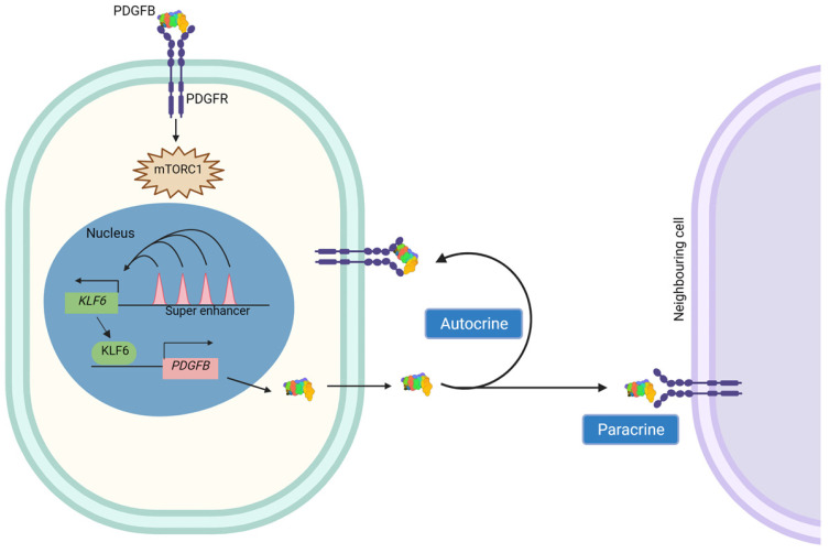 Fig. 1 Full closed-loop signaling schematic for KLF6 super-enhancer mediated PDGFB expression, autocrine/paracrine PDGFR engagement and downstream mTORC1 positive feedback cascade driving pathological pericyte and fibroblast over-proliferation.1
The biological functions of secreted PDGF-BB homodimer subunit are fully focused on PDGFRβ receptor binding and mesenchymal proliferative signal amplification:
Creative Biolabs offers high-quality PDGFB proteins through optimized mammalian secretion systems, including mature PDGF-BB homodimer and isolated receptor-binding domain variants. These products retain native PDGFRβ activation proliferative activity, suitable for pericyte migration and anti-fibrotic inhibitor screening assays. All PDGFB proteins undergo strict quality control to ensure consistent performance and reliable application across diverse stromal research platforms.
Not finding the membrane protein product you need? Contact us to start your one-stop custom service!
Creative Biolabs provides custom-engineered PDGFB stable cell lines, including tumor-derived overexpression and blank empty vector control models. These cell lines are optimized for PDGF growth factor profiling and pericyte migratory signal functional analysis. Each cell line undergoes stringent validation to ensure stable expression profiles during long-term glioma cell culture, and can be deployed for large-scale anti-fibrosis drug screening experiments.
Not finding the stable cell line product you need? Contact us to start your one-stop custom service!
High-specificity recombinant antibodies targeting PDGFB are developed via advanced antibody engineering technologies. These antibodies are validated for platelet and tumor stromal growth factor detection and glioma tissue expression profiling, and can be combined with PDGFRβ detection reagents to analyze complete PDGF-BB receptor signaling complexes in pericyte cell models.
Not finding the recombinant antibody product you need? Contact us to start your one-stop custom service!
Beyond catalog products, Creative Biolabs offers specialized custom services for PDGFB research:
PDGFB forms BB homodimers to activate PDGFRβ and recruit pericytes/fibroblasts for angiogenesis and ECM deposition.
PDGFB drives glioma proliferation and organ fibrosis, a key target for anti-stromal targeted therapy development.
No, all PDGFB products and services are strictly for research use only, not intended for clinical diagnosis or treatment.
Offerings include mature PDGF-BB homodimer proteins, subunit-specific detection antibodies and custom stable cell lines for stromal research.
PDGFB proteins are validated via PDGFRβ binding and pericyte migratory functional testing.








