
Creative Biolabs is offering the most comprehensive services for antibody development projects. With strict regulation and effective execution, we are dedicated to providing the most valuable solutions to complete your projects.
HighlightsCreative Biolabs is offering the most comprehensive services for antibody development projects. With strict regulation and effective execution, we are dedicated to providing the most valuable solutions to complete your projects.
HighlightsCreative Biolabs has established a broad range of platforms for developing novel antibodies or equivalents. These cutting-edge technologies enable our scientists to meet your demands from different aspects and tailor the most appropriate solution that contributes to the success of your projects.
HighlightsWith deep understanding in antibody-related realms and extensive project experience, Creative Biolabs offers a variety of references to help you learn more about our capacities and achievements, including infographic, flyer, case study, peer-reviewed publications, and all kinds of knowledge that can assist your projects. You are also welcome to contact us directly for more specific solutions.
HighlightsGet a real taste of Creative Biolabs, one of the most professional custom service providers in the world. We are committed to providing highly customized comprehensive solutions with the best quality to advance your projects.
Highlights Loading...
All listed services and products are For Research Use Only. Do Not use in any diagnostic or therapeutic applications.
Usherin, the protein product of USH2A gene, is an exceptionally large single-pass transmembrane protein containing 5,202 amino acids in humans. This remarkable protein normally localizes to the periciliary membrane complex (PMC) of photoreceptor inner segments and the ankle link region of cochlear hair cell stereocilia wherein it functions as a critical structural scaffolding component within the Usher interactome. It spends 97% of the protein sequence on its usherin ectodomain and has an extraordinarily high modularity in its extracellular domains, which include one laminin globular-like (LGL) domain, one laminin N-terminal (LN) domain, ten laminin epidermal growth factor-like (LE) repeats, two laminin globular (LG) domains and a whopping 32 fibronectin type III (FN3) domains. These repetitive structural motifs allow usherin to bind multiple times, thereby increasing its potency in binding extracellular matrix competitors (eg, fibronectin and collagen) and transmembrane adhesion receptors. It contains a class I PDZ-binding motif (Asp-Thr-His-Leu) in the intracellular C-terminal tail that directly associates with scaffold proteins whirlin (WHRN) and PDZD7, thus anchoring usherin within the larger USH2 quaternary protein complex that also includes ADGRV1 (GPR98/VLGR1). This functional architecture is required to preserve the mechanical stability of photoreceptor connecting cilia and hair cell mechanosensory bundles. An additional 24 amino acid cochlea-specific splice variant adds to this tissue-specific functional diversification of usherin between exon 70 and 71.
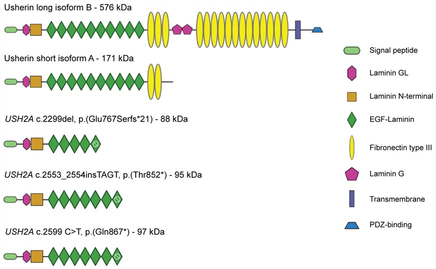 Fig.1 Schematics of usherin domains in the naturally occurring usherin isoforms compared to patient variants and USH2A 13KO.1
For USH2A, the functional repertoire is multi-dimensional across various aspects of sensory neurobiology with a principal homeostasis function in structural maintenance of both photoreceptor and hair cells:
We provide a range of USH2A/usherin protein products and custom constructs designed to support structural and functional research on this challenging target. Due to the large size of full-length usherin and the associated challenges in expression and purification, our portfolio focuses on domain, fragment, and engineered construct formats selected according to research needs and technical feasibility. Available designs may include selected extracellular domains, intracellular regions, and other sequence-defined constructs relevant to USH2A biology. Our recombinant protein platform supports customized construct development and preparation strategies based on individual project requirements. Full-length USH2A, specific expression systems, tags, preparation formats, and functional characterization are confirmed only when supported by project feasibility and corresponding product or validation data.
Not finding the membrane protein product you need? Contact us to start your one-stop custom service!
Establishing cellular models that enable routine and reproducible studies of USH2A variants is challenging, in part because of the large protein size and the requirement for a physiologically relevant expression context. We develop custom-engineered USH2A stable cell models for selected constructs and variants based on project requirements and technical feasibility. These cell lines can support drug screening, protein-protein interaction studies, and functional assays, including applications in high-throughput screening campaigns. Different mammalian host systems and inducible or constitutive expression strategies can be evaluated according to the specific research objective. Cell models carrying selected disease-associated or intronic variants can also be considered on a project-specific basis. Stably transfected cell lines can be characterized using appropriate methods, such as flow cytometry, Western blotting, immunofluorescence localization assays, or functional readouts, with the specific variant, host system, and characterization strategy confirmed for each project.
Not finding the stable cell line product you need? Contact us to start your one-stop custom service!
We provide custom recombinant antibody development for USH2A/usherin to support a range of research applications. Antibody candidates can be developed against selected epitopes or regions of this large protein based on project requirements and antigen accessibility. Target regions, antibody formats, and screening strategies are determined according to the specific research objective and technical feasibility. Developed antibodies may be evaluated for applications such as Western blotting, immunohistochemistry (IHC), immunofluorescence (IF), flow cytometry, or ELISA, depending on the characteristics and performance of individual antibody candidates. Appropriate antibody formats and labeling or conjugation options can also be considered on a project-specific basis to support different experimental workflows.
Not finding the recombinant antibody product you need? Contact us to start your one-stop custom service!
In addition to our catalog offerings, we provide unique custom services in membrane protein and antibody discovery & development. Using our extensive expertise and high-end platforms, we can help with:
No, all reagents and services provided are designated solely for research purposes and cannot be used for diagnostic testing or therapeutic purposes.
Yes. We provide recombinant protein fragments and stable cell lines expressing serial deletion constructs with specified repeat regions. The scaffold of these materials allow mapping the minimal regions required for binding to VLGR1, association with collagen IV, and retention in periciliary membranes.
Yes, these include recombinant protein fragments and stable cell lines expressing known pathogenic truncations and point mutations linked to moderate to sever Usher syndrome type II phenotypes.
Yes we have developed tetracycline-inducible stable lines to express USH2A fragments containing the transmembrane domain and cytoplasmic C-terminal type I PDZ-binding motif. Such configurations allow for both controlled titration of ciliary membrane occupancy, as well as monitoring of whirlin and harmonin recruitment, periciliary membrane extension, and modulation of the phototransduction cascade with temporal precision—avoiding the variability associated with the transient overexpression paradigms used in studying primary cilium biology.








