
Creative Biolabs is offering the most comprehensive services for antibody development projects. With strict regulation and effective execution, we are dedicated to providing the most valuable solutions to complete your projects.
HighlightsCreative Biolabs is offering the most comprehensive services for antibody development projects. With strict regulation and effective execution, we are dedicated to providing the most valuable solutions to complete your projects.
HighlightsCreative Biolabs has established a broad range of platforms for developing novel antibodies or equivalents. These cutting-edge technologies enable our scientists to meet your demands from different aspects and tailor the most appropriate solution that contributes to the success of your projects.
HighlightsWith deep understanding in antibody-related realms and extensive project experience, Creative Biolabs offers a variety of references to help you learn more about our capacities and achievements, including infographic, flyer, case study, peer-reviewed publications, and all kinds of knowledge that can assist your projects. You are also welcome to contact us directly for more specific solutions.
HighlightsGet a real taste of Creative Biolabs, one of the most professional custom service providers in the world. We are committed to providing highly customized comprehensive solutions with the best quality to advance your projects.
HighlightsTo precisely define cell death pathways remains difficult, as apoptosis, necrosis, and metabolic disruption caused by compounds often share similar biochemical traits. In response, we provide a full in vitro service for apoptosis profiling, enabling the specific resolution and quantification of distinct cell death routes. Our system combines several complementary assays, such as Annexin V binding, mitochondrial membrane potential analysis, ROS detection, caspase function mapping, and plasma membrane integrity tests, into an efficient, unified process.
Cellular apoptosis is a genetically programmed and tightly regulated form of cell death essential for development, tissue homeostasis, and immune regulation. Its molecular mechanism involves the activation of cysteine-aspartic proteases through the intrinsic and extrinsic pathways, leading to characteristic morphological and biochemical changes such as membrane blebbing, nuclear fragmentation, and apoptotic body formation. Consequently, performing reliable apoptosis assays is critically important for accurately quantifying cell death, distinguishing apoptosis from necrosis or other forms of cell death, and evaluating the efficacy of therapeutic interventions in immunological research and clinical settings.
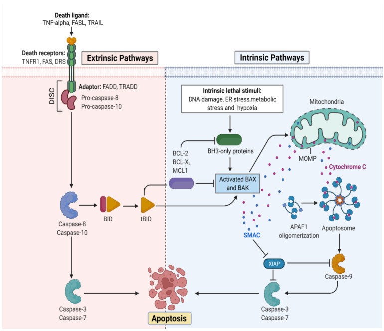 Fig.1 The molecular mechanisms activating intrinsic and extrinsic routes of programmed cell death.1
Creative Biolabs provides a rigorous data analysis platform that converts raw cell death measurements into directly applicable findings for drug development. Whether advancing small-molecule antagonists, antibody-drug conjugates, or CAR-T cell candidates, we offer accurate profiling of apoptotic pathways, enabling a clear distinction between compound-induced metabolic effects and genuine cytotoxic activity.
We offer a homogeneous, add-mix-measure Annexin V staining assay to quantitatively detect externalized phosphatidylserine as an early marker of apoptosis in compound-treated cells using fluorescence plate reader analysis.
We provide a simple mix-add-read JC-10-based assay to evaluate mitochondrial membrane integrity, distinguishing healthy from apoptotic or necrotic cells by measuring red-to-green fluorescence shift.
Our ROS assay enables luminescence-based, add-mix-read quantification of hydrogen peroxide accumulation, facilitating the study of oxidative stress in both apoptotic and necrotic cell death pathways.
We offer a bioluminescent AK leakage assay using minimal cell medium to assess plasma membrane integrity and real-time cytotoxicity kinetics without cell lysis, compatible with multiplexed apoptosis markers.
Our western blot-based service detects proteolytic activation of caspase-3 using an infrared imaging system, providing precise quantification of cleaved versus uncleaved caspase-3 in tumor cell lysates.
We perform a high-throughput-capable, luminescent Caspase 3/7 assay to measure effector caspase activity in a simple add-mix-measure format, enabling robust detection of apoptosis execution across 384-well plates.
Choose our In vitro Cellular Apoptosis Analysis Service for precise, high-content detection of early to late apoptotic markers using distinct assays. We deliver reproducible, statistically robust results with rapid turnaround, supporting your cancer, immunology, or drug development research with expert data interpretation.
To discuss your specific project needs or receive a customized quote for our In Vitro Cellular Apoptosis Analysis Service, please reach out to our scientific team. We are ready to help you generate high-quality, reproducible data for your drug discovery program. Contact us today to start the conversation.
Reference
All listed services and products are For Research Use Only. Do Not use in any diagnostic or therapeutic applications.








