
Creative Biolabs is offering the most comprehensive services for antibody development projects. With strict regulation and effective execution, we are dedicated to providing the most valuable solutions to complete your projects.
HighlightsCreative Biolabs is offering the most comprehensive services for antibody development projects. With strict regulation and effective execution, we are dedicated to providing the most valuable solutions to complete your projects.
HighlightsCreative Biolabs has established a broad range of platforms for developing novel antibodies or equivalents. These cutting-edge technologies enable our scientists to meet your demands from different aspects and tailor the most appropriate solution that contributes to the success of your projects.
HighlightsWith deep understanding in antibody-related realms and extensive project experience, Creative Biolabs offers a variety of references to help you learn more about our capacities and achievements, including infographic, flyer, case study, peer-reviewed publications, and all kinds of knowledge that can assist your projects. You are also welcome to contact us directly for more specific solutions.
HighlightsGet a real taste of Creative Biolabs, one of the most professional custom service providers in the world. We are committed to providing highly customized comprehensive solutions with the best quality to advance your projects.
Highlights Loading...
All listed services and products are For Research Use Only. Do Not use in any diagnostic or therapeutic applications.
Activin A receptor type 1 (ACVR1), also known as activin receptor-like kinase 2 (ALK2), is a approximately 58 kDa type I transmembrane serine/threonine kinase receptor belonging to the transforming growth factor-beta (TGF-β) superfamily. Encoded by the ACVR1 gene on human chromosome 2q23-q24, the mature protein comprises an extracellular ligand-binding domain rich in cysteine residues, a single transmembrane helix, and an intracellular kinase domain with predicted serine/threonine specificity. A distinctive glycine-serine (GS) activation domain juxtamembrane to the kinase domain serves as the critical regulatory switch controlling receptor activation. ACVR1 functions as an indispensable signaling component within bone morphogenetic protein (BMP) receptor complexes. It assembles with type II receptors—including AMHR2, ACVR2A, or ACVR2B—to form heterotetrameric complexes at the cell surface. Upon ligand engagement by BMPs such as BMP7 or GDF2/BMP9, the type II receptor transphosphorylates the GS domain of ACVR1, unleashing its intrinsic kinase activity. Activated ACVR1 subsequently phosphorylates SMAD1/5/8 transcription factors, which translocate to the nucleus to drive gene expression programs governing osteogenesis, chondrogenesis, and tissue patterning. Beyond this canonical BMP pathway, ACVR1 can also engage non-canonical signaling cascades including p38 MAPK and suppress TGF-β/activin signaling under specific contexts, underscoring its signaling versatility.
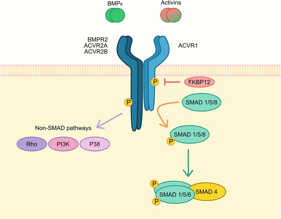 Fig.1 Schematic representation of main ACVR1 signal transduction.1
The biological influence of ACVR1 extends across multiple developmental and pathological systems:
Propel your developmental biology and drug discovery programs with our recombinant ACVR1 membrane protein products. We recognize that the type I receptor topology, GS domain conformational requirements, and ligand-dependent receptor interactions of ACVR1 can present distinct production challenges. To address these, we provide ACVR1 membrane protein preparations and constructs suitable for a range of structural, ligand-binding, biochemical, and functional studies. Available formats and construct designs may vary according to specific research requirements. Each preparation is subject to appropriate quality assessment to support its intended research application.
Not finding the membrane protein product you need? Contact us to start your one-stop custom service!
Reliable, reproducible cellular models are indispensable for dissecting ACVR1-driven biology and evaluating research compounds and pathway modulators. We provide ACVR1 stable cell lines designed to support gain-of-function, loss-of-function, and pathway-focused studies. These platforms can be applied to BMP pathway analysis, SMAD1/5/8 phosphorylation studies, osteogenic differentiation research, and mechanistic or compound screening, providing consistent cellular models for a range of research applications.
Not finding the stable cell line product you need? Contact us to start your one-stop custom service!
Our comprehensive portfolio includes high-affinity recombinant antibodies targeting ACVR1, developed to meet the rigorous demands of various research applications. These antibodies, produced using advanced recombinant technologies, offer superior specificity, sensitivity, and batch-to-batch consistency compared to traditional polyclonal antibodies. Our ACVR1 recombinant antibodies are validated for use in Western Blotting (WB), ELISA, Flow Cytometry (FCM), Immunofluorescence (IF), Immunocytochemistry (ICC), Immunohistochemistry (IHC), and Immunoprecipitation (IP), empowering accurate detection and quantification of ACVR1 in diverse sample types including osteoblast lysates, tumor biopsies, and transfected cell membranes.
Not finding the recombinant antibody product you need? Contact us to start your one-stop custom service!
Beyond our catalog products, we offer specialized custom services for membrane protein and antibody discovery and development. Leveraging our extensive expertise in ACVR1 biology and rare disease pharmacology, we can assist you with:
No, all reagents and services provided are designated solely for research purposes and cannot be used for diagnostic testing or therapeutic purposes.
We can discuss the development of customized co-expression cell models incorporating ACVR1 with BMPR2 or ACVR2A according to specific research requirements. Appropriate expression strategies and pathway-related readouts, such as SMAD1/5/8 phosphorylation or other functional assays, can be selected based on the intended experimental design.
Yes, selected clones have been validated on EDTA-decalcified bone specimens and archival neural tissue following antigen retrieval, yielding specific membranous staining patterns consistent with osteogenic and glial cell distribution. Validation includes peptide competition and recombinant antigen controls to confirm epitope specificity across heterogeneous tissue contexts.
Yes, the immunogen corresponds to a region that is highly conserved across mammalian species, ensuring reliable detection in human, mouse, and rat tissue preparations and cell lysates without requiring multiple species-specific reagents.








