Creative Biolabs is offering the most comprehensive services for antibody development projects. With strict regulation and effective execution, we are dedicated to providing the most valuable solutions to complete your projects.
HighlightsCreative Biolabs is offering the most comprehensive services for antibody development projects. With strict regulation and effective execution, we are dedicated to providing the most valuable solutions to complete your projects.
HighlightsCreative Biolabs has established a broad range of platforms for developing novel antibodies or equivalents. These cutting-edge technologies enable our scientists to meet your demands from different aspects and tailor the most appropriate solution that contributes to the success of your projects.
HighlightsWith deep understanding in antibody-related realms and extensive project experience, Creative Biolabs offers a variety of references to help you learn more about our capacities and achievements, including infographic, flyer, case study, peer-reviewed publications, and all kinds of knowledge that can assist your projects. You are also welcome to contact us directly for more specific solutions.
HighlightsGet a real taste of Creative Biolabs, one of the most professional custom service providers in the world. We are committed to providing highly customized comprehensive solutions with the best quality to advance your projects.
Highlights Loading...
All listed services and products are For Research Use Only. Do Not use in any diagnostic or therapeutic applications.
Bifunctional apoptosis regulator (BFAR) or bifunctional apoptosis inhibitor (BAR), RING finger protein 47 (RNF47) and apoptotic regulatory ring-finger protein, is a multidomain scaffold that converges signaling components from both the extrinsic [death-receptor mediated] and intrinsic [mitochondria-dependent] axes of apopotosis. Recent structural modeling and topology prediction algorithms using AlphaFold, an AI-based approach, show that instead of the one TM domain proposed previously BFAR has four transmembrane segments (TMH1–TMH4) with its N-terminal RING domain positioned on the cytosolic face opposite to the SAM domain. Contrary to earlier speculation, the C-terminal region does not take on a classic DED fold but takes up two pairs of re-entrant helices which contact residues from TMH2 (including S65), TMH3 and TMH4 with high confidence while entering and exiting the membrane from one side. The polytopic membrane topology is required by BFAR to function as an ER-associated E3 ligase, and sets it apart from the cytosolic RING-domain ubiquitin ligases. This protein is expressed in multiple human tissues, most prominently the central and peripheral nerves (note that it may be damaged during postmortem handling of specimen), small intestine, less intensely in liver, skeletal muscle testis as well as immune cells.
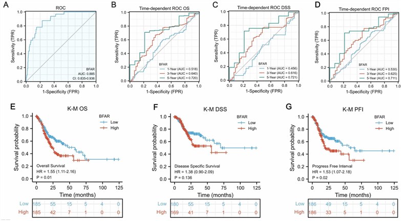 Fig.1 BFAR plays an important role in the diagnosis and prognosis of GC.1
BFAR's functional repertoire spans several axes of apoptosis control, immune signaling, metabolic regulation and disease pathogenesis:
We present a comparison of azole-derivatization-based and bifunctional-reactor-free approaches to engineered cell expression as one proven way that bring an advanced catalogue/set of high-purity BFAR protein products useful for the structure/functional studies/drug research. The use of BFAR is based on our recombinant protein technology platform, enabling BFAR to be produced in a variety of expression systems. Realizing that BFAR is a polytopic membrane protein, it requires proper insertion in the ER to be able to efficiently act as an authentic E3 ligase and interact with substrates; therefore, our portfolio includes full-length BFAR ectodomain with its native four-transmembrane topology as well as carefully crafted constructs suited for specific applications. These can either be detergent-solubilized preparations with mild detergents that maintain its helix bundle structure or membrane-mimetic settings such as nanodiscs, proteoliposomes. Wild-type human BFAR, the deleted RING domain (ΔRING) used to study scaffolding functions without catalytic activity and periphilin-1-like isoform 3 (BFARv3), which is localized in the nucleus with a high level of expression was provided. For informing researchers studying BFAR mediated substrate ubiquitylation, we provide catalytic active and inactive variants for use in vitro reconstitution assays. This scatterplot shows the associated tag configurations both N-terminal His-tag, C-terminal FLAG-tag, Strep-and GST-tags respectively. To ensure suitable performance for your research applications, every batch is quality controlled with various techniques, including SDS-PAGE, Western blot, and SEC-HPLC assessments as well as functional E3 ligase activity assays using PNPLA3 or BI-1.
Not finding the membrane protein product you need? Contact us to start your one-stop custom service!
In light of the central role that BFAR plays in apoptosis regulation, immune signaling, and metabolic disease, developing good cellular models for studying its biology is essential. Custom-engineered BFAR stable cell lines that stably express targets at high levels, including wild-type full-length BFAR, RING domain deleted variants, catalytically inactive RING mutants and periphilin-1-like isoform 3 for assays. These stable cell lines have applications in drug screening, apoptosis assays, TGFβ signaling studies, and Th9 differentiation experiments as well as PNPLA3 degradation assays and high-throughput screening campaigns. Our platform encompasses a range of host backgrounds with inducible or constitutive expression systems. We have generated lines with doxycycline-inducible BFAR expression to study TGFβ signaling, stable knockdown of BFAR for loss-of-function experiments, and expressing complexes between BFAR and PNPLA3 for studies on the mechanisms by which ubiquitylation can influence target protein fate. Stable cell lines each undergo flow cytometry, Western blot, immunofluorescence localisation assays, and functional E3 ligase activity readouts to verify consistent expression was achieved at every passage in a homogenous manner.
Not finding the stable cell line product you need? Contact us to start your one-stop custom service!
We offer a wide range of high-affinity recombinant monoclonal antibodies against BFAR that meet stringent research requirements. Such a design, being based on recombinant technology, allows for a greater specificity, sensitivity, and batch-to-batch consistency compared to the hybridoma-derived antibodies. Because these antibodies are raised again well characterized epitopes spanning both the N-terminal RING domain and SAM (D43-44), as well as C-terminal re-entrant helices near the transmembrane regions in isoforms of BFAR, they can detect full-length ~52 kDa protein along with several possible isoforms. These validated applications include Western blot (confirmed across HEK293, HuH7 & neuronal cell lysates), immunohistochemistry (IHC) of brain, heart and liver tissue sections; as well as IF for ER localization studies, flow cytometry and ELISA. They come in both monoclonal and polyclonal (antibody) formats, which can be conjugated to fluorescent dyes (PE, FITC, Alexa Fluor), enzymes (HRP or AP enzyme-linked antibodies are for ELISA only) or biotin, depending on your experimental workflow.
Not finding the recombinant antibody product you need? Contact us to start your one-stop custom service!
In addition to our catalogue products, we provide unique custom services for the discovery and development of membrane proteins and antibodies. Drawing on classics and world-class platforms, we can help you with:
No— our BFAR products including recombinant proteins, stable cell lines and antibody reagents are strictly limited to non-clinical research only. None has passed the safety, sterility or regulatory threshold necessary before introduction to human subjects.
Yes, we offer two antibodies for each state one that recognizes an epitope masked in the cytosolic resting state but exposed following recruitment into mitochondria (anti-CMN-I) and a second against the C-terminal RING domain present both localization states. Used in parallel, these reagents permit differential immunofluorescence and western blot quantitation of cytoplasmic vs organelle-enriched species, providing a direct measure of stress-induced translocalization without cell fractionation or metabolic labeling approaches.
Yes, stable lines were produced using a doxycycline-regulatable promoter for BFAR transcription. Such an arrangement enables trigger of expression at predefined time points and precise kinetic monitoring of patterning Fas-associated death domain protein interaction, caspase activation attenuation, and survival commitment without the compensatory adaptation artifacts associated for constitutive overexpression systems.
Yes, we license recombinant proteins and stable cell lines expressing established variants with ablated death effector domain heterotypic interaction surfaces or depleted ligases containing zinc-chelating residues. These materials serve as essential specificity controls for separating the contributions of Fas-associated death domain protein sequestration versus caspase-8 inhibition, and ubiquitin chain assembly versus substrate recognition in balancing apoptosis and maintaining cellular homeostasis.