
Creative Biolabs is offering the most comprehensive services for antibody development projects. With strict regulation and effective execution, we are dedicated to providing the most valuable solutions to complete your projects.
HighlightsCreative Biolabs is offering the most comprehensive services for antibody development projects. With strict regulation and effective execution, we are dedicated to providing the most valuable solutions to complete your projects.
HighlightsCreative Biolabs has established a broad range of platforms for developing novel antibodies or equivalents. These cutting-edge technologies enable our scientists to meet your demands from different aspects and tailor the most appropriate solution that contributes to the success of your projects.
HighlightsWith deep understanding in antibody-related realms and extensive project experience, Creative Biolabs offers a variety of references to help you learn more about our capacities and achievements, including infographic, flyer, case study, peer-reviewed publications, and all kinds of knowledge that can assist your projects. You are also welcome to contact us directly for more specific solutions.
HighlightsGet a real taste of Creative Biolabs, one of the most professional custom service providers in the world. We are committed to providing highly customized comprehensive solutions with the best quality to advance your projects.
Highlights Loading...
All listed services and products are For Research Use Only. Do Not use in any diagnostic or therapeutic applications.
Caveolin 2 (CAV2) is a type II single-pass integral membrane structural protein encoded by the CAV2 gene, selectively enriched within plasma membrane caveola invaginations across fibroblast, endothelial and adipocyte cell populations. The polypeptide carries a central hydrophobic membrane embedding domain, short cytoplasmic N-terminal segments and minimal extracellular exposed sequences, and forms hetero-oligomeric assemblies with CAV1 to shape curved caveola membrane microdomains. Caveola compartments function as confined signaling hubs to concentrate surface receptors and small-molecule transporters, and CAV2 participates in maintaining the characteristic curved lipid architecture of these membrane pits while regulating the steady-state residence of signaling complexes within the caveolar space. Without sufficient CAV2 incorporation into oligomeric clusters, caveola structures tend to flatten and lose capacity to retain membrane-bound signal assemblies, which disturbs the ordered transduction of extracellular cues into intracellular response cascades. Different cell subtypes carry distinct basal signaling thresholds, and caveola-dependent buffering relies on balanced CAV2 expression to prevent overamplification of surface receptor activity under resting tissue conditions. Extensive hydrophobic contact surfaces between CAV2 and CAV1 support stable mixed cluster formation, while cytoplasmic binding motifs create temporary docking sites for diverse receptor cytoplasmic tails after ligand engagement.
The sequence variation of CAV2 across the membrane structure domain can interfere with its heterooligomer assembly process and change the basic signal response level of cells in the primary cell model. Although some unrelated lipid-binding proteins can form shallow concave structures in the cell membrane, they can't construct an orderly signal regulation hub, and other caveolin homologous subtypes can't completely replicate the adaptive characteristics and intracellular scaffold function of CAV2 binding CAV1. Under physiological steady-state conditions, CAV2 is stably integrated into the lipid bilayer structure of cell membrane and will not dissociate into soluble cytoplasmic free components. As a type II membrane protein, almost all functional domains of CAV2 are distributed towards cytoplasm, which makes CAV2 have dual functions of reshaping membrane bending structure and instantly anchoring membrane receptor complex, and can accurately regulate the signal conduction intensity of cell membrane. The persistent low expression of CAV2 in cells will lead to the disintegration of pit structure, which will lead to the over-activation of receptor signals on the membrane surface and the imbalance of regulation. Therefore, CAV2 is an important research target to study the signal regulation mechanism of cell membrane microstructure domain.
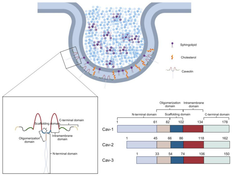 Fig. 1 Schematic of caveola plasma‑membrane microdomain architecture and domain‑organization comparison among caveolin family proteins (Cav‑1, Cav‑2 and Cav‑3), illustrating key functional regions within CAV2.1
The biological functions of integral membrane CAV2 caveolar protein are focused on hetero-oligomer assembly and cytoplasmic signal scaffold binding:
Creative Biolabs offers purified CAV2 membrane protein samples produced under unified preparation workflows, including full-length CAV2 constructs and isolated cytoplasmic interaction domain variants. Truncated fragments cannot support complete hetero-oligomerization and receptor scaffold binding activity, while full-length forms fit caveola microdomain research. All batches receive uniform quality screening. Functional assessments may only be performed under simulated lipid bilayer microenvironment setups. Central membrane embedding and cytoplasmic interaction domain structure is preserved across batches for comparative oligomer testing. Full-length CAV2 membrane samples retain intact inter-caveolin contact surfaces post-purification, supporting reliable detection of transient mixed oligomer complexes in functional comparative analysis.
Not finding the Membrane Protein product you need? Contact us to start your one-stop custom service!
Creative Biolabs provides cell research models with adjustable CAV2 expression levels, suitable for structural observation of caveolar structural proteins and membrane microdomain research. Sample assessment covers sustained caveola membrane detection and CAV1 co-binding analysis, enabling side-by-side comparison of oligomer assembly behavior under varying CAV2 abundances. These cell models can be paired with signal quantification schemes to track receptor activity shifts linked to CAV2 dosage changes.
Not finding the stable cell line product you need? Contact us to start your one-stop custom service!
Anti-CAV2 recombinant antibodies are generated via standardized workflows, compatible with endothelial membrane localization mapping and CAV1-CAV2 heterodimer complex identification. The antibody series supports multi-dimensional visualization of CAV2 distribution within adipocyte and vascular cell compartments.
Not finding the recombinant antibody product you need? Contact us to start your one-stop custom service!
Beyond catalog products, Creative Biolabs offers specialized custom services for CAV2 research:
CAV2 may act as a type II caveolar structural protein to form mixed oligomers with CAV2 and compartmentalize membrane receptor signaling assemblies.
CAV2 expression status might alter caveola architecture and basal receptor signal strength, serving as a key mediator of membrane microdomain homeostasis biological processes.
No, all CAV2 related products and services are strictly for research use only, and cannot be applied in clinical workflows. All material design and functional screening are optimized exclusively for basic laboratory research, without meeting clinical application criteria.
Offerings include full-length CAV2 membrane protein, target-specific recombinant antibodies and tunable expression cell research models, supporting research on caveola assembly and compartmentalized signaling.
Laboratory analysis schemes may include co-oligomer binding assays to assess heterodimer formation capacity under simulated lipid bilayer environments.








