
Creative Biolabs is offering the most comprehensive services for antibody development projects. With strict regulation and effective execution, we are dedicated to providing the most valuable solutions to complete your projects.
HighlightsCreative Biolabs is offering the most comprehensive services for antibody development projects. With strict regulation and effective execution, we are dedicated to providing the most valuable solutions to complete your projects.
HighlightsCreative Biolabs has established a broad range of platforms for developing novel antibodies or equivalents. These cutting-edge technologies enable our scientists to meet your demands from different aspects and tailor the most appropriate solution that contributes to the success of your projects.
HighlightsWith deep understanding in antibody-related realms and extensive project experience, Creative Biolabs offers a variety of references to help you learn more about our capacities and achievements, including infographic, flyer, case study, peer-reviewed publications, and all kinds of knowledge that can assist your projects. You are also welcome to contact us directly for more specific solutions.
HighlightsGet a real taste of Creative Biolabs, one of the most professional custom service providers in the world. We are committed to providing highly customized comprehensive solutions with the best quality to advance your projects.
Highlights Loading...
All listed services and products are For Research Use Only. Do Not use in any diagnostic or therapeutic applications.
Chitinase 3-like 1 (CHI3L1), was originally characterized as a secreted serum glycoprotein with molecular weight of approximately 40kDa and is classified in the glycoside hydrolase family18(GH18). It binds to chitin polysaccharides such as oligomers and polymers but does not have intrinsic enzyme activity to cleave polymerized chains. Originally purifying this unique protein from MG63 osteosarcoma cell line supernatant, it has now been established as a master regulator of almost every organ system capable for tissue damage/inflammation/repair/remodeling. CHI3L1 is abundantly expressed by macrophages, neutrophils, chondrocytes, synoviocytes and vascular smooth muscle cells (VSMC), as well as in astrocytes but its expression can be significantly increased under conditions of type 2 immune activation, tissue damage or cellular stress. CHI3L1 is composed of a conserved-barrel fold, typical for metabolic enzymes, containing a short N-terminal signal peptide that directs secretion followed by the catalytic domain and ending with an optional carbohydrate-binding motif (CBM) or chitin binding domain to allow specific high-affinity interactions between Chi3l1 proteins and their effectors including: chitooligosaccharides heparin collagen. CBM carries essential tryptophan residues that dramatically change the conformation of CBM upon ligand binding, and histidine 209 acts as a chitin-sensing residue promoting innate immune responses in close proximity to sites where invading organisms are likely found such as fungi (the fungal cell wall polymer chitin) or even nematodes. Although it does not possess the six cysteine residues necessary for bona fide chitinase activity, CHI3L1 retains the CBM and acts as a lectin-like signaling molecule that can bind various cell surface receptors such as receptor for advanced glycation end products (RAGE), syndecan-1/αVβ3 integrin, interleukin-13 receptor alpha 2 (IL-13Rα2) and vascular endothelial growth factor receptor 2 (VEGFR2).
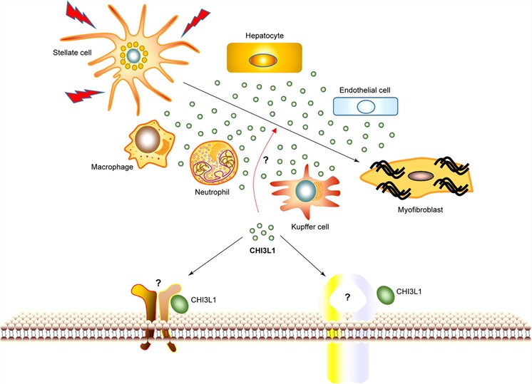 Fig.1 Possible effects of CHI3L1 on liver fibrosis.1
The functional repertoire of CHI3L1 is broad, encompassing multiple facets innate immunity and wound healing; tumor biology; neuroinflammation:
We provide a comprehensive range of premium CHI3L1 protein products required for structural, functional, and therapeutic antibody development research. We have an in-house recombinant protein technology to produce CHI3L1 expression systems such as mammalian cells (Expi293F, HEK293, CHO), insect cells, and even bacterial hosts. As CHI3L1 is highly glycosylated, a secreted protein for which receptor binding requires the proper folding of its α/β-barrel domain, our portfolio specifically focuses on full-length CHI3L1. Available formats encompass Avitag- and His-tagged proteins for biotinylation and affinity purification, tag-free proteins relevant to X-ray crystallography and structural studies, as well as C-terminal deletion mutants that allow the dissection of carbohydrate-binding motif function. Available wild-type human CHI3L1, H209A mutant to probe chitin–sensing activity and shelf-stable variants. Characterized CHI3L1 proteins for rabbit immunization, epitope binning in the identification of binding sites and humanizing antibodies are readily available to researchers interested in antibody development. All batches are quality controlled via SDS-PAGE, Western blot and SEC-HPLC together with heparin affinity chromatography as well as functional receptor binding assays to ensure optimal applicability for your experiments.
Not finding the protein product you need? Contact us to start your one-stop custom service!
It's a fundamental step for investigating CHI3L1, especially due to the important role of this protein in many diseases such as inflammation, cancer or neurodegeneration. Here we present high-expression CHI3L1 stable cell lines (wild-type, CBM deletion mutants and secretion-deficient variants) that have been custom-engineered for you. They are also optimal for drug screening, macrophage polarization assays and angiogenesis tube formation studies, neuroinflammation model systems (e.g. co-culture models) or high-throughput screening campaigns. Our cell line development platform accommodates a wide variety of host backgrounds such as HEK293, CHO-K1, and several cancer/normal cells, we have created doxycycline (Dox)-inducible CHI3L1 expression lines for controlled inflammation studies; stable CHI3L1 knockdown (KD) lines for loss-of-function analyses and lastly reporter-expressing cell-lines now allowing us to monitor secretion dynamics of secretory proteins. Flow cytometry, Western blot, immunofluorescence localization assays and functional cytokine induction readouts are used to validate each stable cell line (passed separately) for consistency and homogeneity of expression over passages.
Not finding the stable cell line product you need? Contact us to start your one-stop custom service!
We provide a complete portfolio of recombinant antibodies against CHI3L1, which have high-affinity and fulfill the rigorous requirements for research. Generated with contemporary recombinant technologies, these antibodies typically allow for more specificity and sensitivity as well as improved batch-to-batch consistency than hybridoma-derived ones. We raised our anti-CHI3L1 antibodies against carefully designed epitopes across the N-terminal region, α/β catalytic barrel, 4α–5β loop, and 4α helix (the functional epitope identified for blocking Abs), as well as C-terminal CBM to ensure a strong detection of the full-length 40 kDa protein. Applications that have been validated include WB, IHC of tumor and liver sectioning respectively, IF for localization studies in astrocyte and macrophage tissues sections where CHI3L1 was present compared with controls, flow cytometry to analyze the expression levels measured by qPCR or IL-8 compared to countercontrols (unstimulated samples) were also using ELISA functional blocking assays based on previously demonstrated role of angiogenesis/inflammation mediated through this molecule. Available in both monoclonal and polyclonal formats, with conjugation to fluorescent dyes (PE, FITC, Alexa Fluor), enzymes (HRP, AP) or biotin, depending on your workflow.
Not finding the recombinant antibody product you need? Contact us to start your one-stop custom service!
In addition to our catalog products, we provide custom services for protein and antibody discovery & development. With our rich experience and advanced platforms, we can help you with:
No, all the CHI3L1 products and services are for research use only (RUO) and not intended to be used in clinical diagnosis, prevention or treatment or cure of any disease.
For example, we have created stable line in which CHI3L1 transcription is controlled by a tetracycline-responsive promoter. Such a system permits precise temporal triggering of expression at defined time points with progressive CD206 upregulation, collagen deposition and extracellular matrix remodeling tracked over multiple intervals without the chronic activation biases associated with constitutive overexpression platforms.
Depending on the specific clone and supporting datasheet, selected anti-CHI3L1 antibodies may be suitable for immunofluorescence analysis in appropriate fixed-cell or tissue-based research samples. Application conditions, sample compatibility, and staining performance should be determined according to the product-specific validation data and experimental design.
Yes, the protein is in a known, buffered endotoxin free buffer at physiologic pH that enables TIM-barrel folding and glycan-mediated stabilization for lectin activity. This enables direct dilution into pull-down reactions, biolayer interferometry sensorgrams or vapour diffusion crystallisation setups without buffer exchange from the supplied formulation.








