
Creative Biolabs is offering the most comprehensive services for antibody development projects. With strict regulation and effective execution, we are dedicated to providing the most valuable solutions to complete your projects.
HighlightsCreative Biolabs is offering the most comprehensive services for antibody development projects. With strict regulation and effective execution, we are dedicated to providing the most valuable solutions to complete your projects.
HighlightsCreative Biolabs has established a broad range of platforms for developing novel antibodies or equivalents. These cutting-edge technologies enable our scientists to meet your demands from different aspects and tailor the most appropriate solution that contributes to the success of your projects.
HighlightsWith deep understanding in antibody-related realms and extensive project experience, Creative Biolabs offers a variety of references to help you learn more about our capacities and achievements, including infographic, flyer, case study, peer-reviewed publications, and all kinds of knowledge that can assist your projects. You are also welcome to contact us directly for more specific solutions.
HighlightsGet a real taste of Creative Biolabs, one of the most professional custom service providers in the world. We are committed to providing highly customized comprehensive solutions with the best quality to advance your projects.
Highlights Loading...
All listed services and products are For Research Use Only. Do Not use in any diagnostic or therapeutic applications.
Ectonucleoside triphosphate diphosphohydrolase 1 (ENTPD1), commonly known as CD39, is a broadly distributed membrane-anchored ectoenzyme of the E-NTPDase family. ENTPD1 is abundantly expressed on the surface of regulatory T lymphocytes, tumor-associated macrophages (TAMs), myeloid-derived suppressor cells (MDSC) and vascular endothelium and at least one subset of activated B-cells. Its main biochemical role is in the ordered hydrolysis of extracellular ATP and ADP to AMP, thus removing inflammatory danger signals from the microenvironment while simultaneously providing substrate for 5'-ectonucleotidase CD73–oligomerization produces a novel cyclodextrin-like structure which promotes cell-to-cell adhesion or inductive signaling. This enzymatic relay converts ATP-enriched immunostimulatory microenvironment into adenosine-rich immune-suppressive environment that triggers the A2A and A2B receptors on effector T cells and natural killer (NK) to inhibit degranulation, cytokines secretion, and proliferation. ENTPD1 was significantly upregulated in the malignant context across non-small cell lung cancer, colorectal adenocarcinoma, melanoma and ovarian carcinoma where it formed a powerful metabolic barrier to anti-tumor immunity that positively correlated with tumor resistance mediated by programmed death 1 (PD-1) blockade. This convergence of biological and clinical knowledge has raised the profile for ENTPD1 as a leading therapeutic target in which monoclonal antibodies and catalytic inhibitors are progressing through Phase I/II trials, next-generation immuno-oncology agents.
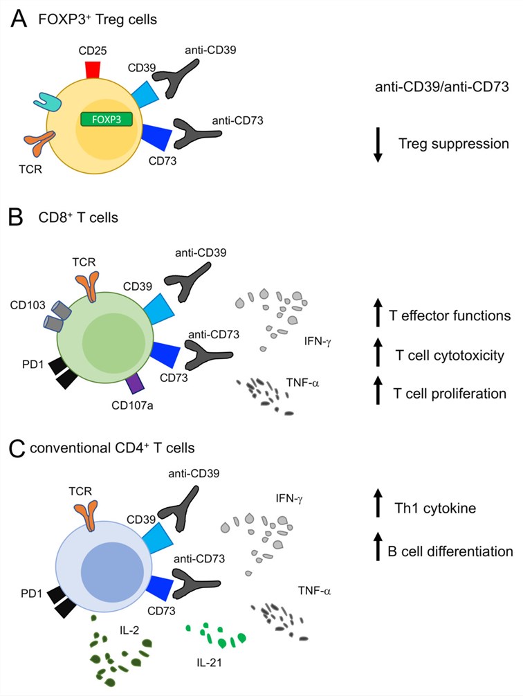 Fig.1 Effects of targeting CD39 on T cells.1
ENTPD1 runs multiple biological programs suggestive of more than being just a simple nucleotide hydrolase:
We have developed a portfolio of ENTPD1 membrane protein products to support purinergic enzymology, structural biology, binding studies, and antibody research. Because ENTPD1 is an ectonucleotidase with membrane-spanning regions and a catalytic extracellular domain, appropriate protein format and structural integrity are important considerations for relevant experimental applications. We provide ENTPD1 constructs and preparations suitable for enzyme activity studies, structural characterization, ligand or inhibitor binding assays, and related functional research. Available formats and construct designs may vary according to specific research requirements, with appropriate quality assessment applied based on the intended application.
Not finding the membrane protein product you need? Contact us to start your one-stop custom service!
The generation of cellular models that accurately recapitulate native ENTPD1 biology has been critical to disentangling purinergic immune suppression and accelerating therapeutic development. We have developed a strong panel of stable cell lines based on lentiviral delivery and transposon-mediated genome engineering, which enable authentic presentation at the plasma membrane, expression of native glycosylation moieties and significant ectonucleotidase activity in context to inflammatory stimuli under hypoxic enrichment. In addition to native constructs, our platforms feature catalytically inactive mutants containing mutations at the ACR4 coordination site; polymorphisms that have naturally occurred in human populations and affect enzyme kinetics or signaling outputs (e.g. responses of adenosine receptors); and coexpression systems with an engineered full-length adenosinergic axis by co-expressing ENTPD1/CD73/adenosine A2A receptor/P2X7 R) within a single cellular background. We also constructed reporter-coupled lines for identifying conditions of strong adenosine-mediated immunosuppression in real time after ENTPD1 inhibition or substrate challenge, with both cAMP response element-driven luciferase and NFAT promoter elements. Complementing this, loss-of-function (in principle) models generated via shRNA-mediated knockdown or dominant-negative expression are available for rescue testing and synthetic lethality screening. Genomic integration mapping, surface enzyme activity profiling, and functional testing in T-cell suppression co-cultures and ATP hydrolysis/adenosine generation assays comprise stringent validation for every cell line. The well-defined and scalable cellular substrates provide essential tools to study the mechanism of immuno-oncology, anti-tumor compound screening, as well as in pre-clinical optimization of biologics targeting the ENTPD1 axis.
Not finding the stable cell line product you need? Contact us to start your one-stop custom service!
Through our proprietary monoclonal antibody engineering platform we have designed a focused family of sequence-defined high-affinity monoclonal antibodies for each investigational target area to help facilitate the study of this ectonucleotidase immune checkpoint including specificity determinations required in order to navigate the structural complexities associated with ENTPD1 investigation. These clones are selective for the substrate-occupancy of catalytically active ENTPD1, recognizing both glycosylation-dependent epitopes in the extracellular catalytic domain and conformational determinants at within a specific membrane-proximal stalk region; and they discriminate ENTPD1 from paralogs (ENTPD2, ENTPD3) with negligible cross-reactivity. These antibodies are thoroughly characterized across quantitative Western blotting under reducing and non-reducing conditions, direct and competitive ELISA formats for soluble ENTPD1 detection in biological fluids, flow cytometric staining of live tumor-infiltrating leukocyte and endothelial populations, high-resolution immunofluorescence/immunocytochemical confocal microscopy imaging of immunological synapses/vessel junctions to determine subcellular patterns/polarity/localization within environmental niches such as tumors or lymphoid tissues where engaged T-cells influence growth factor stimulation at an endpoint level compared to diglycerides (HLA-A,TGF-β) surrounding immune-mediate localization/inflammation/early vascular response signaling network function per-tumor microvascular niche type; pancrelipase–membrane transduction pathway modulators/signaling networks associated overall functional effects on receptor-mediated signal desensitization between ligand-bound receptors pre/post-different cytokine infusion locations relative each other identified based cetuximab-ligand mediated EGRF pathways.
Not finding the recombinant antibody product you need? Contact us to start your one-stop custom service!
In addition to our standard catalog, we provide full-scale definition and engineering capabilities specifically for ENTPD1:
Certainly. We often generate catalytically inactive versions containing mutations in the ACR4 coordination motif, glycosylation-site mutants affecting ectodomain stability and naturally occurring polymorphisms associated with altered enzyme kinetics. They are provided either as purified recombinant proteins or genetically stable cell lines with complete sequencing, mass spectrometric identity confirmation and functional characterizations by substrate hydrolysis and phosphate release assays.
Yes. You're in mammalian expression systems, which guarantee folding of the extracellular catalytic domain and proper N-linked glycosylation while maintaining divalent cation-binding sites necessary for ATP and ADP hydrolysis. Functional integrity of each batch is confirmed prior to release using analytical verification via malachite green phosphate release, HPLC-based nucleotide substrate turnover and surface plasmon resonance for binding studies.
Yes. We can discuss customized co-expression cell models incorporating ENTPD1 with CD73 or selected purinergic receptors according to specific research requirements. Cell model design, expression strategy, and appropriate readouts for nucleotide metabolism, adenosine signaling, or related pathway studies can be determined based on the intended experimental design.
Absolutely. The monoclonal antibodies that we raised were directed against distinct epitopes of the ENTPD1 ectodomain and displayed limited sequence homology to the other family members' (ENTPD2,11; ENTPD3,9–13; ENTPD5 or 8). Their exquisite specificity, as confirmed by extensive cross-reactivity testing using Western blot, ELISA and flow cytometry according to a standardized protocol developed for their use may provide diagnostic quantification of these complex conformational antibodies from challenging biological compartments including serum/plasma/tumor digests.








