
Creative Biolabs is offering the most comprehensive services for antibody development projects. With strict regulation and effective execution, we are dedicated to providing the most valuable solutions to complete your projects.
HighlightsCreative Biolabs is offering the most comprehensive services for antibody development projects. With strict regulation and effective execution, we are dedicated to providing the most valuable solutions to complete your projects.
HighlightsCreative Biolabs has established a broad range of platforms for developing novel antibodies or equivalents. These cutting-edge technologies enable our scientists to meet your demands from different aspects and tailor the most appropriate solution that contributes to the success of your projects.
HighlightsWith deep understanding in antibody-related realms and extensive project experience, Creative Biolabs offers a variety of references to help you learn more about our capacities and achievements, including infographic, flyer, case study, peer-reviewed publications, and all kinds of knowledge that can assist your projects. You are also welcome to contact us directly for more specific solutions.
HighlightsGet a real taste of Creative Biolabs, one of the most professional custom service providers in the world. We are committed to providing highly customized comprehensive solutions with the best quality to advance your projects.
Highlights Loading...
All listed services and products are For Research Use Only. Do Not use in any diagnostic or therapeutic applications.
Mucosal vascular addressin cell adhesion molecule 1 (MADCAM1; also known as mucosal addressin cell adhesion molecule-1) is a single-pass type I transmembrane glycoprotein of the immunoglobulin superfamily that is expressed selectively at postcapillary venules in the endothelium of mucosal and lymphoid tissues. The architecture of human MADCAM1 supports its bifunctional role in the step-wise process that leads to lymphocyte transendothelial migration. The extracellular region (residues 19–317) contains two N-terminal immunoglobulin-like domains, D1, an IgV-type domain, and D2, an IgC2-type domain, that collectively constitute the integrin-binding interface mediating α4β7-dependent adhesion. Just distal to D2 is an O-glycosylated serine/threonine-rich mucin-like domain with a negatively charged stalk that raises the Ig domains above the endothelial glycocalyx and can carry sialyl-Lewis X carbohydrates involved in L-selectin (CD62L)-mediated lymphocyte rolling. A membrane-proximal Ig-like domain present in some MADCAM1 isoforms differs between species. The protein has a single transmembrane helix (residues 318–338) embedded in the plasma membrane and a short cytoplasmic region (residues 339–382) involved in membrane-associated intracellular interactions.
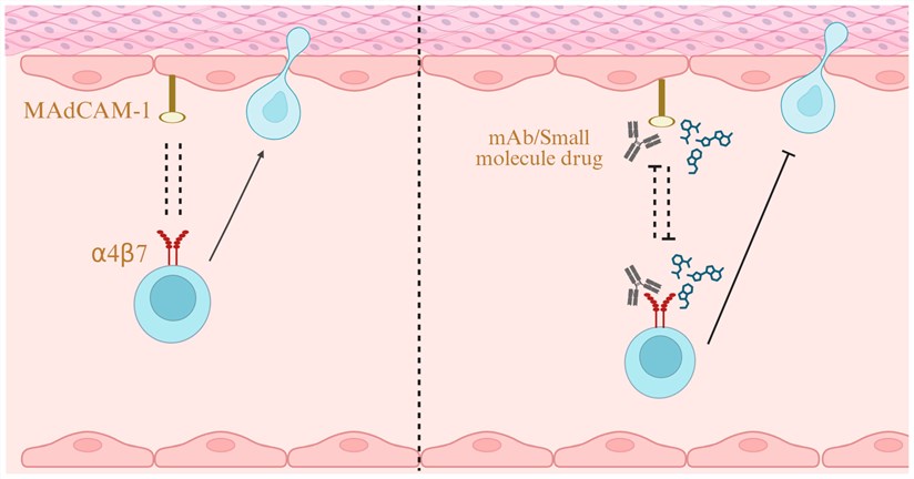 Fig.1 α4β7/MAdCAM-1 as a Drug Target.1
MADCAM1 has a multidimensional functional repertoire spanning mucosal immunology, lymphocyte trafficking, and the pathogenesis of inflammatory disease:
We provide a range of high-quality MADCAM1 protein products to support structural and functional studies of this mucosal adhesion receptor. Because the extracellular region of MADCAM1 contains immunoglobulin-like and mucin-like domains with important post-translational modifications, appropriate construct design and protein format are important considerations for relevant research applications. Our products can support structural studies, α4β7-related binding research, antibody development and characterization, and other studies of MADCAM1-mediated cell adhesion. Available construct formats, tags, and other product characteristics should be determined according to the corresponding product information.
Not finding the protein product you need? Contact us to start your one-stop custom service!
Our custom-engineered MADCAM1 stable cell lines in 6 different recipient backgrounds using cell-based clones are available on request. The stable cell lines that you develop will be suitable for drug screening, lymphocyte adhesion assays under shear flow conditions, T cell costimulation studies and high-throughput screen campaigns. Our cell line development platform covers a variety of host backgrounds like HEK293, CHO-K1 or even endothelial cell lines such as HUVEC with either inducible or constitutive expression systems. We have established lines from which stable doxycycline-regulated MADCAM1 expression can be produced, enabling precise control over lymphocyte homing; derived other lines to express MADCAM1 on the apical surface of polarized epithelial monolayers for transmigration assays; and yet others where stable depletion of endogenous MAdCAM-1 enables mechanistic studies. Both flow cytometry, Western blot, and immunofluorescence localization assays are performed on each stable cell line to ensure consistent expression throughout passage as well as functional lymphocyte adhesion readouts.
Not finding the stable cell line product you need? Contact us to start your one-stop custom service!
Our portfolio includes recombinant antibodies targeting MADCAM1, designed to support a range of research applications. Recombinant production provides sequence-defined, renewable reagents with consistent lot-to-lot production. Depending on the specific antibody and supporting datasheet, selected MADCAM1 antibodies may be suitable or available for applications such as Western Blotting, Immunohistochemistry (IHC), Immunofluorescence (IF), ELISA, and other antibody-based assays. These antibodies can support the detection and analysis of MADCAM1 in appropriate research samples and model systems. Product-specific specificity, affinity, species reactivity, epitope information, conjugation options, and application compatibility should be determined according to the corresponding antibody data. For specialized research needs, customized MADCAM1 recombinant antibody discovery and development can also be discussed according to the intended experimental design.
Not finding the recombinant antibody product you need? Contact us to start your one-stop custom service!
In addition to our catalog products, we provide dedicated custom services for membrane protein and antibody discovery & development. With a proven history and our cutting-edge platforms, we can help you with:
We provide various kinds of recombinant proteins including soluble, membrane-tethered format wild-type or domain-deleted MADCAM1 expression clones; also stable cell lines expressing proper human and mouse formatted full-length or Fc chimera with corresponding quality controls.
Mammalian expression system (HEK293, CHO, NS0) to achieve appropriate O-glycosylation of mucin domain for L-selectin binding and full biological activity. It is learned that Fc chimera proteins can be produced in NS0 myeloma cells and with native glycosylation. Ig domain fragments without the mucin region may also be expressed in insect cell systems.
Yes, selected clones were tested and are indeed positive on archival gastrointestinal specimens after antigen retrieval; they stain specifically along high endothelial venules in the lamina propria and with variable expression overlying follicle-associated epithelium of Peyer's patches. Further validation consists of peptide competition and integrin-blocking absorption controls to validate epitope specificity in addition to ligand-occupation dependence among heterogeneous tissue contexts.
Yes, we have produced dual-stable lines that constitutively express full-length MADCAM1 at one locus and integrin alpha4beta7 from another independent locus. We report a release of each batch with documentation for shear-independent binding, as well as transpiring kinetics between adhesive and stiff states within the parallel-plate flow chamber, which allows for standardized cellular platforms that exist without transfecting every time before mechanistic and therapeutic antibody screenings.








