Creative Biolabs is offering the most comprehensive services for antibody development projects. With strict regulation and effective execution, we are dedicated to providing the most valuable solutions to complete your projects.
HighlightsCreative Biolabs is offering the most comprehensive services for antibody development projects. With strict regulation and effective execution, we are dedicated to providing the most valuable solutions to complete your projects.
HighlightsCreative Biolabs has established a broad range of platforms for developing novel antibodies or equivalents. These cutting-edge technologies enable our scientists to meet your demands from different aspects and tailor the most appropriate solution that contributes to the success of your projects.
HighlightsWith deep understanding in antibody-related realms and extensive project experience, Creative Biolabs offers a variety of references to help you learn more about our capacities and achievements, including infographic, flyer, case study, peer-reviewed publications, and all kinds of knowledge that can assist your projects. You are also welcome to contact us directly for more specific solutions.
HighlightsGet a real taste of Creative Biolabs, one of the most professional custom service providers in the world. We are committed to providing highly customized comprehensive solutions with the best quality to advance your projects.
Highlights Loading...
All listed services and products are For Research Use Only. Do Not use in any diagnostic or therapeutic applications.
MAGE family member A4 (MAGEA4) is a Type I cancer-testis antigen (CTA), defined by an M age homology d omain of approximately 200 amino acids and molecular weight about 35 kDa. In contrast to proteins that are broadly expressed and used for housekeeping, MAGEA4 is one of the most tissue-commissioned physiological-expressed antigens that retains expression mainly in germ cells within the seminiferous tubules of males testes where it has been identified as an antigen candidate plus several differentially which seem like placental trophoblasts but little. In light of these characteristics, with aberrant reactivation in a wide oncological spectrum from urothelial carcinoma to esophageal squamous cell carcinoma and non-small-cell lung cancer (NSCLC) through melanoma as well as multiple gastrointestinal cancers by the concepts of oncoimmunology their restricted normal-tissue expression pattern makes MAGEA4 one of the most UNESCO attractive targets. MAGEA4 localizes intracellularly in the cytoplasm and nucleus while which is involved with key protein-protein interactions with tumor suppressor pathways to inhibit apoptosis such as p53 and HDACs, allowing malignant proliferation to continue. In addition, MAGEA4-derived peptides can be presented on human leukocyte antigen (HLA) class I molecules which are recognized by cytotoxic T lymphocytes confirming that the development of adoptive T-cell receptor therapies and therapeutic cancer vaccines along with antibody-based precision oncology in humans is built upon evidence supporting a role for this important cornerstone tumor-associated antigens. Due to its tumor-selective expression profile, direct oncogenic functionality and peptides that are exceptionally immunogenic.
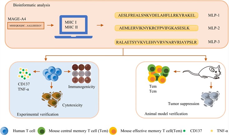 Fig.1 Schematic diagram of the screening procedure for the MAGE-A4 long peptides(MLPs).1
MAGEA4 orchestrates many of these pathological and therapeutically exploitable programs as more than just a tumor-associated antigen:
We provide full-length MAGEA4 proteins designed to support structural biology, antibody development, and related biochemical research. Because proper folding of the MAGE homology domain is important for structural and epitope-focused studies, appropriate expression and protein preparation strategies can be selected according to specific experimental requirements. Available full-length MAGEA4 proteins and engineered constructs can support studies of protein structure, antibody binding, protein interactions, and related research applications.
For studies of antigen presentation and T-cell receptor recognition, MAGEA4-derived peptide–HLA complexes should be considered separately from full-length MAGEA4 protein. These peptide–HLA reagents can be used to investigate TCR–pMHC interactions, binding properties, and related immune-recognition mechanisms when an appropriate MAGEA4-derived peptide and HLA allele are selected. Product format, analytical characterization, and functional suitability should be determined according to the specific reagent and supporting product data.
Not finding the protein product you need? Contact us to start your one-stop custom service!
Developing cellular models that most accurately reproduce the native expression patterns of MAGEA4 and its tissue-specific subcellular localization will be essential in elucidating the oncogenic mechanisms through which it operates as well as for assessing immunotherapeutic approaches. Harnessing lentiviral transduction with the uniquely advantageous properties of a recently developed MAR minicircle genome integration technology, we established an array of MAGEA4 stable cell lines characterized by physiological expression levels, normal nuclear-cytoplasmic partition and faithful responses to genotoxic/inflammatory stimuli. In addition to wild-type MAGEA4, our cellular platform also encompasses loss-of-function truncation mutants, phosphorylation-site variants with known effects on protein stability as well its interaction partners p53 and HDAC1. We also provide HLA-restricted reporter systems, with luciferase and fluorescent markers downstream of NF-κB, p53 or interferon-stimulated response elements that can measure antigen presentation only in real-time since T-cell engagement is not measured directly but rather by measuring transcription from genes turned on through innate immune activation. Quality assurance procedures for each cell line include genomic integration verification by digital droplet PCR, assessment of expression stability at extended passage numbers and functional validation in MAGEA4-specific siRNA knockdown rescue experiments.
Not finding the stable cell line product you need? Contact us to start your one-stop custom service!
We have established an in-depth and diverse antibody repertoire for MAGEA4, developed explicitly to facilitate precision oncology analytics and companion diagnostic development. These sequence-defined monoclonal antibodies (mAbs) specifically recognize native and denatured MAGEA4 conformations, discriminate between the closely related MAGE family paralogs, including isoforms such as phosphorylated or ubiquitinated variants of the protein, which is relevant to a more complete ontology for studies into molecular evolution with implications in cancer. They include: quantitative Western blotting (both under reducing and non-reducing conditions), direct/sandwich ELISA for detection of soluble MAGEA4 in tissue lysates and biological fluids, flow cytometric determination of the staining characteristics with permeabilized versus unpermeabilized tumor cell lines vs. primary patient samples; high-resolution immunofluorescence-based confocal microscopy to analyze targeting specific nuclear/cytoplasmic compartments better than other complementary protein assays will explore.
Not finding the recombinant antibody product you need? Contact us to start your one-stop custom service!
Far exceeding our traditional catalog, we provide comprehensive discovery and manufacturing solutions specifically designed around the demands of MAGEA4-targeted researches:
Absolutely. We generated our monoclonal antibodies against novel epitopes that exist within the specific regions of MAGEA4 immediately flanking this highly conserved homology domain. Comprehensive cross-reactivity analyses against recombinant MAGEA1, MAGEA3, and the immunosuppressive-derived human cancer antigens (HCA) proteins together with some individual parallel studies of protein lysate stability over time provided remarkable specificity well suited for diagnostic purpose in biomarker quantification either by using non-denaturing or denaturating-based methods such as Western Blots.
Absolutely. The monoclonal antibodies we raised against unique epitopes in the MAGEA4-specific regions proximal and distal to the homology domain conserved amongst all members of this family. Tested for extensive cross-reactivity against hundreds of recombinant MAGEA1, MAGEA3 and 10 proteins (rMAGE) as well as more than a dozen other related proteins have revealed exquisite specificity making these reagent suitable reagents capable to be employed in diagnostic immunohistochemistry combined with quantification biomarkers even from complex tissue lysates.
Yes. Our proprietary mammalian and insect cell expression systems—as well as our in-house refolding protocols designed for the bacterial-derived material— guarantee that MAGE homology domain can form its native α-helical bundle structure. The conformational integrity of each batch is confirmed by circular dichroism spectroscopy and functional relevance to established TCR clones for HLA class I peptide binding as well as tetramer staining assays.
Certainly. We regularly create site-directed mutants in the MAGE homology domain, N-and C-terminal truncation constructs and rare phosphomimetic variants to probe phosphorylation-dependent stability and interaction dynamics. These custom constructs come as purified recombinant proteins or genetically stable cell lines, and a comprehensive data packet that includes full sequence verification and functional characterization.