
Creative Biolabs is offering the most comprehensive services for antibody development projects. With strict regulation and effective execution, we are dedicated to providing the most valuable solutions to complete your projects.
HighlightsCreative Biolabs is offering the most comprehensive services for antibody development projects. With strict regulation and effective execution, we are dedicated to providing the most valuable solutions to complete your projects.
HighlightsCreative Biolabs has established a broad range of platforms for developing novel antibodies or equivalents. These cutting-edge technologies enable our scientists to meet your demands from different aspects and tailor the most appropriate solution that contributes to the success of your projects.
HighlightsWith deep understanding in antibody-related realms and extensive project experience, Creative Biolabs offers a variety of references to help you learn more about our capacities and achievements, including infographic, flyer, case study, peer-reviewed publications, and all kinds of knowledge that can assist your projects. You are also welcome to contact us directly for more specific solutions.
HighlightsGet a real taste of Creative Biolabs, one of the most professional custom service providers in the world. We are committed to providing highly customized comprehensive solutions with the best quality to advance your projects.
Highlights Loading...
All listed services and products are For Research Use Only. Do Not use in any diagnostic or therapeutic applications.
NPHS1 (nephrin) is a single pass transmembrane adhesion molecule encoded by NPHS1 gene. This protein localizes to specialized plasma membrane regions that constitute intercellular junction structures within particular tissue types. NPHS1 carries multiple extracellular immunoglobulin like domains, a transmembrane segment and a long intracellular cytoplasmic region capable of assembling multi‑molecular molecular complexes. Distinct from signalling oriented receptors, its primary molecular activities centre on intercellular contact rather than triggering strong downstream signal cascades upon molecular binding. This molecule accumulates at narrow intercellular contact interfaces where multiple adhesion associated molecules assemble together to build highly organized membrane architecture. Additional auxiliary membrane anchored molecules co‑localize within the same junction compartments and can cooperate to stabilise local molecular arrangements. Spatial distribution across cell surface is tightly controlled, and the molecule tends to concentrate within defined membrane subdomains instead of spreading evenly across entire plasma membrane.
Proper assembly of intercellular junction architecture depends on coordinated intermolecular contacts among multiple membrane anchored adhesion molecules. Changes in NPHS1 surface abundance could disturb the arrangement of local inter‑cell contact interfaces, which might modify barrier related tissue properties. Altered NPHS1 molecular structure appears to correlate with disrupted tissue junction organization, making this molecule a valuable subject for cell junction focused research. Other adhesion molecules cannot fully replicate the structural contributions supplied by NPHS1 within its native tissue niche. Variations of NPHS1 surface presentation may contribute to reorganized intercellular contact patterns within tissue microenvironments. Tissue barrier properties emerge from collective behaviours of multiple adhesion components, so perturbation of one molecular component may shift overall interface characteristics without fully eliminating barrier function. Different physiological states may drive subtle adjustments to NPHS1 abundance and spatial arrangement, supporting adaptive tuning of intercellular contact interfaces according to local tissue requirements.
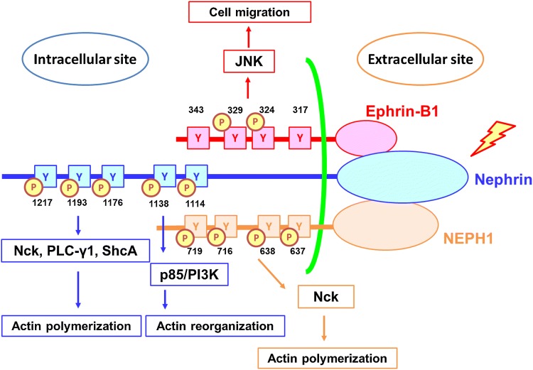 Fig. 1 Schematic representation of NPHS1 transmembrane signaling architecture. Following intracellular‑domain tyrosine phosphorylation, NPHS1 recruits multiple cytoplasmic adaptor molecules, which further mediate actin‑cytoskeleton polymerization and reorganization events.1
The biological functions of NPHS1 are focused on intercellular molecular adhesion, junction associated complex assembly and tissue barrier structural organization:
Creative Biolabs offers purified NPHS1 membrane samples via standardized preparation workflows, including full‑length NPHS1 constructs and isolated extracellular structural variants. Isolated extracellular fragments may not support complete junction related complex assembly behaviours, while full‑length constructs may be suited for intercellular adhesion and molecular scaffold assembly related research. All samples receive routine quality screening, and functional relevant observation may only be carried out with full‑length samples under simulated membrane environments. All sample batches follow unified processing standards to maintain consistent structural features for comparative laboratory analysis across separate test groups.
Not finding the membrane protein product you need? Contact us to start your one-stop custom service!
Creative Biolabs provides adjustable NPHS1 expression cell models with varied expression levels, applicable to transmembrane adhesion molecule structural characteristic observation and inter‑cell contact related research. Sample evaluation includes sustained target expression detection and preliminary intermolecular interaction associated observation, which can support comparative analysis of adhesion related behaviours under different expression statuses. These cell systems can be matched with diverse laboratory analysis schemes to observe changes of intermolecular contact efficiency under different target expression abundances.
Not finding the stable cell line product you need? Contact us to start your one-stop custom service!
Antibody reagents targeting NPHS1 are generated via mature protein preparation workflows, compatible with multiple routine laboratory detection methods for cellular localization profiling and molecular complex identification, to support systematic analysis of NPHS1 distribution and junction associated molecular complexes across diverse laboratory research setups. The antibody series can cooperate with other common laboratory detection reagents to complete multi dimensional observation of target distribution inside tissue samples.
Not finding the recombinant antibody product you need? Contact us to start your one-stop custom service!
Beyond catalog products, Creative Biolabs offers specialized custom services for NPHS1 research:
NPHS1 may function as a transmembrane adhesion molecule and participate in intercellular contact and junction associated multi‑molecular complex assembly.
NPHS1 expression status may alter intercellular junction architecture and tissue barrier associated properties, serving as a major regulatory mediator of cell junction related biological processes.
No, all NPHS1 related products and services are strictly for research use only, not intended for clinical related operations. All material designs and functional tests are only optimized for basic laboratory research scenarios, without matching clinical application standards.
Offerings include full‑length NPHS1 membrane protein, target specific recombinant antibodies and adjustable expression cell research models, supporting cell junction and tissue barrier related molecular research.
Laboratory observation schemes may include adhesion related intermolecular contact tests to analyse molecule associated behaviours under simulated cellular environments.








