
Creative Biolabs is offering the most comprehensive services for antibody development projects. With strict regulation and effective execution, we are dedicated to providing the most valuable solutions to complete your projects.
HighlightsCreative Biolabs is offering the most comprehensive services for antibody development projects. With strict regulation and effective execution, we are dedicated to providing the most valuable solutions to complete your projects.
HighlightsCreative Biolabs has established a broad range of platforms for developing novel antibodies or equivalents. These cutting-edge technologies enable our scientists to meet your demands from different aspects and tailor the most appropriate solution that contributes to the success of your projects.
HighlightsWith deep understanding in antibody-related realms and extensive project experience, Creative Biolabs offers a variety of references to help you learn more about our capacities and achievements, including infographic, flyer, case study, peer-reviewed publications, and all kinds of knowledge that can assist your projects. You are also welcome to contact us directly for more specific solutions.
HighlightsGet a real taste of Creative Biolabs, one of the most professional custom service providers in the world. We are committed to providing highly customized comprehensive solutions with the best quality to advance your projects.
Highlights Loading...
All listed services and products are For Research Use Only. Do Not use in any diagnostic or therapeutic applications.
Ribosomal protein SA (RPSA; also called 40S ribosomal protein SA) is a multifunctional 295-amino acid–long and poilerasome12833-9-diameter, danilo. Its architecture is typically characterized by two major domains: an N-terminal domain homologous to prokaryotic ribosomal protein S2 (RPS2) that adopts a folded globular structure with α/β fold, and the intrinsically disordered C-terminal domain, highly conserved in vertebrates. RPSA has exceptional structural flexibility: the N-domain is partially folded, while the C domain was found to be intrinsically disordered, and full-length RPSA can exist in several folding states that may be required for its functional roles within multiple cellular localizations. RPSA is an acylation modification at ser2 with fatty acids post-translationally affecting which appears to contribute to the establishment of interactions between lipid membrane, and so it may represent a mechanism that controls on the accession from two different oligomeric forms, comparing monomers and dimers. However, this protein does not undergo glycosylation and is phosphorylated at Tyr139, which can be cleaved by stromelysin-3 (ST3/MMP11) on the cell surface. RPSA is constitutively expressed in all human tissues with greatest expression noted within the ovary (RPKM 874.9), thymus (RPKM 760.4), and many other organs as it serves a crucial role as part of the core component that associates with rRNA into maintaining ribosomal structure within eukaryotic cells. RPSA is known to be localized in multiple cellular compartments: co-precoding with 40S ribosomal subunit as a structural component; associated protein of chromatin and histone – nuclear localization, perinuclear / DNA localization that may connect the potential link from nuclear envelope (NE) to chromatin DNA; cell surface receptor for laminin-laminin related proteins — extracellular ligands.
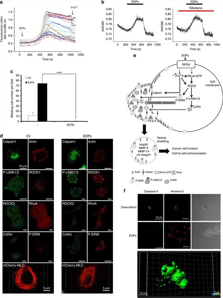 Fig.1 EDP-induced blebbing depends on calcium influx and on RhoA/ROCK/MLC signalling pathway.1
RPSA has a remarkably plastic structure and conserved evolutionarily characteristics with functional repertoire:
Creative Biolabs offers a portfolio of RPSA recombinant proteins designed to support research in cancer biology, ribosome biology and ribosomopathy research, and host–pathogen interactions. Our protein products may include RPSA constructs from different species and in different recombinant formats according to product availability and experimental requirements. These proteins can support applications such as laminin-binding studies, investigation of ribosome-related functions, pathogen-interaction research, antibody development and characterization, and compound screening.
Not finding the protein product you need? Contact us to start your one-stop custom service!
Cell lines are valuable tools for investigating RPSA biology in a controlled cellular context. Creative Biolabs offers custom-engineered RPSA stable cell lines designed to support consistent target expression or regulation according to specific research requirements. These models can support studies of laminin binding, ribosome-related functions, host–pathogen interactions, cellular mechanisms, and compound-response screening. RPSA overexpression or loss-of-function models may also be developed for research on laminin-mediated adhesion, cell migration, and other RPSA-associated processes. Expression strategies, reporter systems, functional readouts, and characterization methods should be selected according to the specific project design and supporting data. These cell models are intended for research use only.
Not finding the stable cell line product you need? Contact us to start your one-stop custom service!
With our extensive portfolio of high-affinity recombinant antibodies against RPSA, we have developed antibody products that fulfill the highest requirements for research applications. Unlike traditional polyclonal antibodies, which consist of a mixture of different antibody molecules isolated from serum or tissue extracts that have the potential to show inconsistency between batches, these are produced using modern recombinant technologies and exhibit superior specificity, sensitivity and batch-to-batch consistency. The RPSA recombinant antibodies are validated in-house and can be used for WB, ELISA, FCM, IF, ICC/IHC/ISH/IP on many different sample types, such as: tumor biopsy, colon carcinoma tissue; lung cancer cell lysates, and prion-infected brain homogenates. serum samples. Antibodies that differentiate the 37-kDa monomeric (37LRP) versus 67-kDa dimeric (67LR) forms of RPSA allow identification and precise characterization of its maturation status, both physiologically in embryonic cell lineages such as ES cells or pathophysiologically infiltrating cancer stem-like/transitional neoplastic tumor propagating units. Further N-terminal (residues 161–179; laminin-binding domain), C-terminal intrinsically disordered domain, and the acylation site at Ser2 reagents allow for more refined investigations of RPSA structure-function relationships and post-translational modification states.
Not finding the recombinant antibody product you need? Contact us to start your one-stop custom service!
On top of our catalog portfolio, Creative Biolabs provides custom protein engineering and antibody discovery solutions targeting the specific needs associated with RPSA-centric research programs:
No, all RPSA products and services are designated as research use only. They are not intended to make clinical diagnoses, or for therapeutic purposes.
Yes, the protein is concentrated in a defined, magnesium-containing, endotoxin-monitored buffer at physiological pH that preserves both ribosome-binding and laminin-recognition surfaces. This supports immediate addition to reticulocyte lysate translation reactions, coupling to laminin-coated matrices, or sedimentation velocity experiments without dialysis or buffer exchange.
Yes, we provide antibodies to human recombinant proteins and stable cell lines expressing pathogenic substitutions within the laminin-binding propeller or ribosomal interface. These materials to provide a platform for direct comparison of 40S assembly efficiency, laminin-1 dissociation constants and surface presentation kinetics relative along wild-type benchmarks in controlled cellular backgrounds.
Yes, selected clones were validated on methanol-fixed and paraformaldehyde-fixed preparations to provide discrete nucleolar, diffuse cytoplasmic, or peripheral punctate staining patterns that co-localize with established fibrillarin (nucleolus), ribosomal protein S6 (cytoplasm) and vinculin (periphery). Competitions with peptides and ribosome-dissociation controls are performed to validate epitope accessibility in the different states.








