
Creative Biolabs is offering the most comprehensive services for antibody development projects. With strict regulation and effective execution, we are dedicated to providing the most valuable solutions to complete your projects.
HighlightsCreative Biolabs is offering the most comprehensive services for antibody development projects. With strict regulation and effective execution, we are dedicated to providing the most valuable solutions to complete your projects.
HighlightsCreative Biolabs has established a broad range of platforms for developing novel antibodies or equivalents. These cutting-edge technologies enable our scientists to meet your demands from different aspects and tailor the most appropriate solution that contributes to the success of your projects.
HighlightsWith deep understanding in antibody-related realms and extensive project experience, Creative Biolabs offers a variety of references to help you learn more about our capacities and achievements, including infographic, flyer, case study, peer-reviewed publications, and all kinds of knowledge that can assist your projects. You are also welcome to contact us directly for more specific solutions.
HighlightsGet a real taste of Creative Biolabs, one of the most professional custom service providers in the world. We are committed to providing highly customized comprehensive solutions with the best quality to advance your projects.
Highlights Loading...
All listed services and products are For Research Use Only. Do Not use in any diagnostic or therapeutic applications.
SPG7 matrix AAA peptidase subunit, paraplegin (SPG7) is also known as paraplegin, CAR 140257 or SPG5C. It contains a multi-pass transmembrane structure where it functions as an ATP-dependent metalloprotease protein with about 795-amino acid and calculated molecular weight of approximately 88.6 kDa. It contains two hydrophobic transmembrane helices (TM1, residues 145–165; TM2, residues 249–269) that flank an intermembrane space (IMS) domain with well-structured secondary and tertiary structure. The IMS region is composed of an α-helix, three β-strands (β1–β3), two short α-helices (α1–α2), and a connecting β-strand (β4) with the arrangement resulting in an independent folding element between these two transmembrane helices that can stabilize the m-AAA protease hexameric complex. The AAA+ domain contains characteristic Walker A and B motifs, Sensor 1 and 2 motifs as well as a pore loop (IGG) that lines the central channel of the hexameric ring to define substrate specificity. The catalytically active zinc-binding site essential for proteolytic activity is found within the C-terminal metallopeptidase domain. SPG7 forms a complex with its paralogous partner AFG3L2 (ATPase family gene 3-like 2) in the mitochondrial inner membrane where it assembles to create m-AAA protease, an ATP-dependent protein quality control hetero-oligomeric complex affording hexameric ring structure of 900 kDa. Besides its canonical protease activity, SPG7 was recently shown to be a conserved and essential constituent of the mitochondrial permeability transition pore (PTP), an inner membrane channel that opens at contact sites between outer and inner membranes. In this role, SPG7 interacts with cyclophilin D (CypD) and voltage-dependent anion channel (VDAC), modulating Ca2+- and ROS-induced pore opening, mitochondrial membrane potential collapse, leading to cell death. SPG7 expression level is highest in the thyroid (RPKM 11.5), skin (RPKM 10.1) and is expressed across all human tissues, reiterating its essential role for mitochondrial homeostasis alongside much ubiquitously expressed genes.
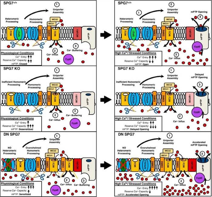 Fig.1 Central hypothesis. Schematic of m-AAA protease processing and regulation of MCU in the mitochondria.1
SPG7 has a complex molecular architecture that reflects its functional contributions to mitochondrial proteostasis, membrane dynamics and neuronal protection:
Creative Biolabs provides a highly purified portfolio of SPG7 membrane and protease research preparations for functional and structural studies of this mitochondrial AAA protease. Our Membrane Protein technology supports the development of SPG7 protein formats tailored to different experimental requirements. These preparations can support applications such as ELISA, antibody production and characterization, biochemical studies, protein interaction research, and investigations of mitochondrial quality-control mechanisms. They offer researchers useful tools for studying interactions between SPG7 and associated proteins such as AFG3L2, CypD, VDAC, or protein substrates. Specific protein formats, complex assembly strategies, structural characteristics, and functional assays are selected and confirmed according to the corresponding project requirements and available experimental data.
Not finding the membrane protein product you need? Contact us to start your one-stop custom service!
Generating stable cellular models is paramount for SPG7 research. Creative Biolabs is capable of delivering custom-designed SPG7 membrane protein stable cell lines to achieve high and consistent expression levels of target SPG7. These stable cell lines can be used for drug screening, ATPase activity assays, protease activity assays or PTP opening assay, as well as mitochondrial Ca²⁺ retention studies and high-throughput screening thus providing a uniform platform to study SPG7 biology and pharmacology in vitro. We work carefully to ensure optimal expression and cellular viability, streamlining your research workflow. We comprise both SPG7-overexpressing lines (cell models for investigating m-AAA complex assembly and PTP regulation) and SPG7-knockdown lines with validated expression verified by flow cytometry, Western blotting, and functional ATPase activity assays. The ability to use specialized reporter cell lines, with mitochondrial membrane potential sensors (TMRE, JC-1) or Ca2+ indicators (Rhod-2, mt-GCaMP), that allow real-time monitoring of PTP dynamics makes them very useful for performing therapeutic screening in neurodegeneration and ischemia-reperfusion injury models.
Not finding the stable cell line product you need? Contact us to start your one-stop custom service!
Combining all these factors, we present you with a wider portfolio of high-affinity recombinant antibodies against SPG7 that can meet your experimental needs. These high-quality antibodies are generated through recombinant technologies, which provide higher specificity, sensitivity and batch-to-batch consistency than traditional polyclonal antibodies. SPG7 recombinant antibodies are validated for in vitro use in applications such as Western Blotting (WB), ELISA, Flow Cytometry (FCM), Immunofluorescence (IF); ICC, IHC and IP with the potential to detect SPG7 from various types of samples, including muscle biopsies, fibroblast lysates, mitochondrial preparations, as well as cerebrospinal fluid and formalin-fixed paraffin-embedded tissue sections. We provide antibodies that bind to specific SPG7 epitopes — the N-terminal FtsH domain for complex assembly studies, the AAA+ ATPase domain involved in enzyme activity assays, and the C-terminal metallopeptidase domain mapping of catalytic residues as well as IMS region SLiM searching, providing multiple points at which it is possible to render anatomical fidelity with respect to SPG7 across a broad range of health disease signs and following therapeutic intervention.
Not finding the recombinant antibody product you need? Contact us to start your one-stop custom service!
In addition to our catalog products, Creative Biolabs provides high-throughput custom services for membrane protein and antibody discovery & development:
No, all Creative Biolabs' SPG7 products and services are for research use only and not to be used in clinical diagnosis, prevention treatment or cure of any disease.
Yes, we offer complex-selective Abs that detect conformational epitopes on SPG7 when it is hetero-hexamerized with AFG3L2 and inserted into the inner membrane.
Yes, we have created dual-stable lines of cells containing constitutive expression from independent loci using both full-length SPG7 and AFG3L2. Individual batches are supplied with functional assays of mitochondrial membrane potential maintenance, OPA1 cleavage product ratios and cristae junction density yielding a defined cell due to unrestrained mechanistic studies without transient transfection.
Yes, some clones have been validated on fixed and permeabilized cells using it to compare against mitochondrial markers with punctate staining patterns co-localising COX4 TOM20. The staining is enriched in subsarcolemmal and intermyofibrillar mitochondrial aggregates on skeletal muscle cryosections.








