
Creative Biolabs is offering the most comprehensive services for antibody development projects. With strict regulation and effective execution, we are dedicated to providing the most valuable solutions to complete your projects.
HighlightsCreative Biolabs is offering the most comprehensive services for antibody development projects. With strict regulation and effective execution, we are dedicated to providing the most valuable solutions to complete your projects.
HighlightsCreative Biolabs has established a broad range of platforms for developing novel antibodies or equivalents. These cutting-edge technologies enable our scientists to meet your demands from different aspects and tailor the most appropriate solution that contributes to the success of your projects.
HighlightsWith deep understanding in antibody-related realms and extensive project experience, Creative Biolabs offers a variety of references to help you learn more about our capacities and achievements, including infographic, flyer, case study, peer-reviewed publications, and all kinds of knowledge that can assist your projects. You are also welcome to contact us directly for more specific solutions.
HighlightsGet a real taste of Creative Biolabs, one of the most professional custom service providers in the world. We are committed to providing highly customized comprehensive solutions with the best quality to advance your projects.
Highlights Loading...
All listed services and products are For Research Use Only. Do Not use in any diagnostic or therapeutic applications.
Tumor necrosis factor superfamily member 10 (TNFSF10), also known as TRAIL (TNF-related apoptosis-inducing ligand), Apo-2 ligand (Apo-2L, CD253 or TL2) has a pectic structure with a complete length of 281 amino acids and is categorized as tumor necrosis factor (TNF)20 ligand superfamily. Biological activity requires this trimeric architecture, enabling a single TNFSF10 trimer to simultaneously bind three receptor molecules, forcing the receptors into clusters that are respectively much more effective than bivalent agonistic antibodies in activating cell death. The most unique biological feature is its ability to preferentially induce apoptosis in transformed and tumorous cells but leaving most normal cells relatively unharmed; this tumor-selective cytotoxicity has gained TNFSF10 a position as one of the best new biotherapeutic agents against cancers. Such selectivity is dependent on the expression of TNFSF10 receptor types, cancer cells generally express death receptors (DR4/TRAIL-R1 and DR5/TRAIL-R2) that have functional death domains whereas normal cells upregulate decoy receptors that lack a death domain to sequester ligand without undergoing apoptosis (DcR1/TRAIL-R3 and DcR2/TRAIL-R4). TNFSF10's roles extend beyond oncology into immune surveillance, inflammation, tissue remodeling, cardiovascular protection and with emerging roles in the pathogenesis of Alzheimer's disease and the regulation of autoimmunity.
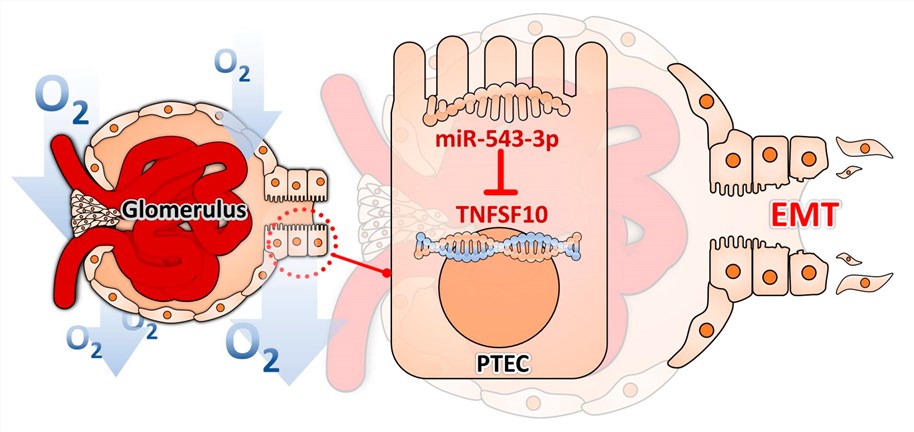 Fig.1 Illustration of the mechanism by which hypoxia induced EMT in PTECs through miR-545-3p–TNFSF10.1
Its functional diversity in apoptosis induction, immune modulation, and tissue homeostasis:
Creative Biolabs offers a comprehensive selection of high-purity recombinant TNFSF10 proteins designed to support cancer immunology and apoptosis research. Our protein expression platforms provide TNFSF10 in research-ready formats suited to different experimental requirements. These well-characterized proteins can support receptor-binding studies, apoptosis-related assays, drug screening, and mechanistic investigations of extrinsic apoptotic signaling. Specific species, oligomeric formats, tags, quality-control specifications, and available bioactivity data are provided according to the corresponding product datasheet or individual project requirements.
Not finding the membrane protein product you need? Contact us to start your one-stop custom service!
The establishment of strong cellular models is essential for TNFSF10 research. Creative Biolabs is able to provide custom-engineered TNFSF10 stable cell lines expressing high levels of target TNFSF10 protein that will reliably reproduce your assay conditions. This stability makes these cell lines the best choice for drug screening, ligand binding studies, apoptosis assays, DISC formation assays, and high-throughput screening, providing a more stable and reproducible platform for studying TNFSF10 biology and pharmacology. We ensure maximum expression with cellular integrity, expediting your research pipeline. TNFSF10-overexpressing lines for studying death receptor activation and immune cell-mediated cytotoxicity were created, as were TNFSF10-knockdown lines to investigate loss-of-function effects. Following transduction, expression is validated by flow cytometry, Western blotting, and functional apoptosis assays. Developing DR5-dependent luciferase or caspase-8 activation readouts in specialized reporter cell lines allows real-time monitoring of extrinsic apoptotic signaling, making them powerful tools for research screening in cancer and autoimmune disease models.
Not finding the stable cell line product you need? Contact us to start your one-stop custom service!
Whether you're focused on immunotherapy or other disciplines of research, we offer recombinant antibodies with high-affinity binding to TNFSF10 that can stand up to the stringent requirements today in the lab. Developed leveraging novel recombinant technologies, these antibodies offer superior specificity, sensitivity and batch-to-batch consistency over polyclonal counterparts. TNFSF10 recombinant antibodies have been tested with WB, ELISA, FC, IF and have been demonstrated to be suitable for use with serum (e.g. human), plasma (e.g. human), and various sample types including cell culture supernatants, tumor biopsies and immune cell lysates enabling the accurate detection and quantification of TNFSF10 proteins in multiple subcellular compartments used for biological study. We supply antibodies that differentiate between membrane-bound and soluble TNFSF10 isoforms, one targeting the receptor-binding domain for inhibition assays, as well as other that recognizes only the N-terminal cytoplasmic tail (for subcellular localization). Alternative agents recognizing post-translationally-modified TNFSF10 (tyrosine-phosphorylated by PKDCC/VLK) enable complex studies of ligand-activation states.
Not finding the recombinant antibody product you need? Contact us to start your one-stop custom service!
In addition to our extensive catalog offerings, Creative Biolabs also offers customized solutions for protein engineering and antibody discovery dedicated to the unique needs of your TNFSF10 research programs:
No, Creative Biolabs TNFSF10 products and services are strictly intended for research use only.
Yes. We generated phospho-state-selective antibodies that recognize the serine-phosphorylated epitope within the N-terminal regulatory region revealed upon hypoxia-inducible factor stabilization, but masked following phosphatase-mediated deactivation.
Yes. In such dual-stable lines, we have expressed the pro-autophagic protein BNIP3 together with either a directly mitochondrial-targeted fluorescent protein or an LC3-based autophagosome reporter from independent expression cassettes.
Yes. Optimized clones have been confirmed on formalin-fixed specimens after a heat-mediated antigen retrieval, resulting in specific perinuclear and punctate cytoplasmic staining patterns that are enhanced towards necrotic tissue cores and co-localize with established mitochondrial markers.








