
Creative Biolabs is offering the most comprehensive services for antibody development projects. With strict regulation and effective execution, we are dedicated to providing the most valuable solutions to complete your projects.
HighlightsCreative Biolabs is offering the most comprehensive services for antibody development projects. With strict regulation and effective execution, we are dedicated to providing the most valuable solutions to complete your projects.
HighlightsCreative Biolabs has established a broad range of platforms for developing novel antibodies or equivalents. These cutting-edge technologies enable our scientists to meet your demands from different aspects and tailor the most appropriate solution that contributes to the success of your projects.
HighlightsWith deep understanding in antibody-related realms and extensive project experience, Creative Biolabs offers a variety of references to help you learn more about our capacities and achievements, including infographic, flyer, case study, peer-reviewed publications, and all kinds of knowledge that can assist your projects. You are also welcome to contact us directly for more specific solutions.
HighlightsGet a real taste of Creative Biolabs, one of the most professional custom service providers in the world. We are committed to providing highly customized comprehensive solutions with the best quality to advance your projects.
HighlightsCancer remains a significant global health challenge. Currently, immune checkpoint inhibitors (ICIs) lead to advancements in cancer immunotherapy. Targeting co-inhibitory immune checkpoint molecules has shown promising results in treating cancers like melanoma and non-small cell lung cancer. Normally, these molecules help regulate immune responses by inhibiting cytotoxic T-cell activity, thus preventing autoimmunity. However, in cancer, they allow tumors to evade immune detection. Hence, targeting these inhibitory pathways holds substantial potential for effective cancer treatment.
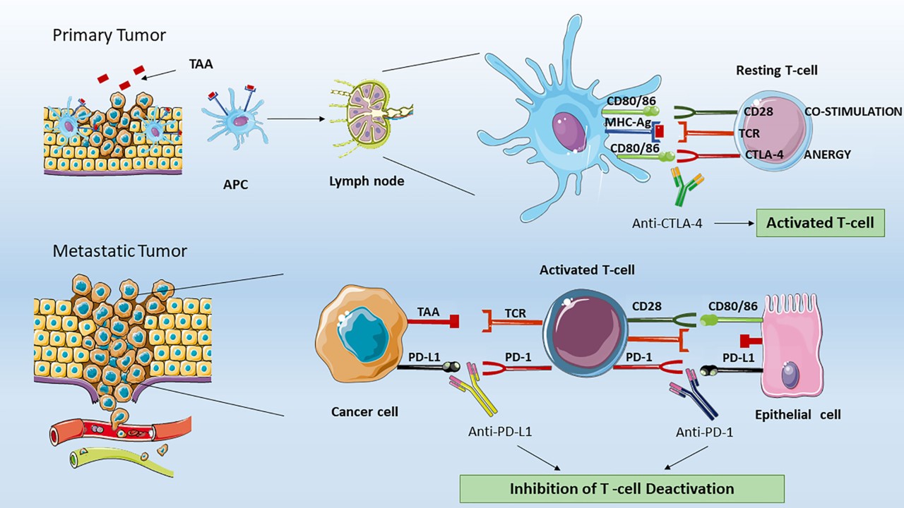 Fig.1 Various ICIs have different effects on T lymphocytes.1, 3
Creative Biolabs offers cutting-edge inhibitory immune checkpoint-targeted reporter gene assay to assess the efficacy of therapeutic compounds. Our cell-based reporter gene assay employed top-tier inhibitory immune checkpoint-expressing reporter cell lines that are engineered or optimized with effective reporter systems, providing quantitative readouts for thorough analysis after treatment. At the same time, we offer ready-to-use reporter cell line products and sensitive report plasmids for global customers to operate in your labs. In addition, we also specialize in tailoring unique solutions to fit your exact research needs. With a skilled team of experts, we guarantee high-quality result delivery with a short turnaround of just 2 weeks.
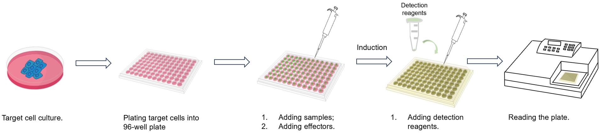 Fig.2 Workflow of our assay.
The success of inhibitory immune checkpoint targeted therapies in cancer treatment has exerted a positive influence on the research of these checkpoints. Therefore, Creative Biolabs delivers a full range of inhibitory IC-targeted reporter assays to support researchers around the world in discovering and developing more effective targeted therapeutics and therapies. Listed below are several popular targeted reporter gene assays, including:
We also provide various engineered reporter plasmids for global customers, enabling seamless integration into diverse research projects. Meanwhile, we offer a series of services such as codon optimization and sequence optimization to construct or optimize customized reporter plasmids in accordance with customers' unique research purposes.

| Summary | This study developed a PD-L1 bioassay using engineered Jurkat T cells (Jurkat/PD-1/NFAT-Luc) and CHO cells expressing TCRA and PD-L1. The bioassay aimed to evaluate the anti-PD-L1 activity of an anti-PD-L1/CD40L bispecific antibody fusion protein (anti-PD-L1/CD40L BsAbFP). Cells were exposed to varying concentrations of anti-PD-L1/CD40L bispecific antibody or anti-PD-L1 monoclonal antibody. Both treatments activated Jurkat/PD-1/NFAT-Luc cells in a dose-dependent manner, demonstrating that the anti-PD-L1 arm of the bispecific antibody fusion protein effectively promotes T cell activation, comparable to the anti-PD-L1 mAb. |
| Sample Type | Anti-PD-L1/CD40L Bispecific Fusion Protein |
| Result: |
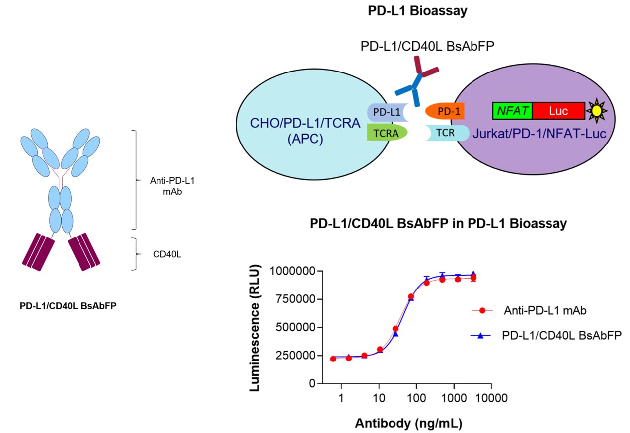 Fig.4 The structure of anti-PD-L1/CD40L BsAbFP (Left) and activity evaluation of anti-PD-L1 arm (Right).2, 4 |
For more information on our inhibitory immune checkpoint-targeted reporter gene assay, feel free to contact us.
References
All listed services and products are For Research Use Only. Do Not use in any diagnostic or therapeutic applications.








