
Creative Biolabs is offering the most comprehensive services for antibody development projects. With strict regulation and effective execution, we are dedicated to providing the most valuable solutions to complete your projects.
HighlightsCreative Biolabs is offering the most comprehensive services for antibody development projects. With strict regulation and effective execution, we are dedicated to providing the most valuable solutions to complete your projects.
HighlightsCreative Biolabs has established a broad range of platforms for developing novel antibodies or equivalents. These cutting-edge technologies enable our scientists to meet your demands from different aspects and tailor the most appropriate solution that contributes to the success of your projects.
HighlightsWith deep understanding in antibody-related realms and extensive project experience, Creative Biolabs offers a variety of references to help you learn more about our capacities and achievements, including infographic, flyer, case study, peer-reviewed publications, and all kinds of knowledge that can assist your projects. You are also welcome to contact us directly for more specific solutions.
HighlightsGet a real taste of Creative Biolabs, one of the most professional custom service providers in the world. We are committed to providing highly customized comprehensive solutions with the best quality to advance your projects.
Highlights Loading...
All listed services and products are For Research Use Only. Do Not use in any diagnostic or therapeutic applications.
CACNA1G (Calcium voltage‑gated channel subunit alpha1 G) encodes a multi‑pass transmembrane T‑type low‑voltage‑activated calcium‑channel pore‑forming α‑subunit predominantly localised to plasma‑membrane compartments, with minor protein fractions detected within intracellular vesicular membrane structures.This channel subunit occurs across multiple organ‑derived tissue populations and displays distinct, tissue‑biased expression profiles, presenting detectable abundance within excitable tissue groups including nervous‑system‑related compartments.Distinct from soluble intracellular polypeptides, it possesses four repeated structural domains, each containing six transmembrane helical segments, alongside conserved extracellular pore‑forming loops and extensive cytoplasmic terminal segments harbouring interaction motifs for downstream partner engagemen.It functions as the pore-forming α1G subunit of the CaV3.1 channel and does not require the auxiliary subunits that are characteristic of high-voltage-activated CaV1/CaV2 channel complexes. Small membrane depolarizations activate the channel from relatively negative potentials, opening a Ca2+-selective pore and generating a transient inward T-type Ca2+ current. Rapid voltage-dependent inactivation limits the duration of Ca2+ entry, while channel recovery and reactivation can contribute to repetitive electrical activity in excitable cells.
Sequence-level alterations in CACNA1G can modify channel gating, activation and inactivation kinetics, or Ca2+ permeability and thereby alter voltage-dependent Ca2+ entry. CaV3.1 is one of three T-type channel isoforms, alongside CaV3.2 and CaV3.3, and is encoded by CACNA1G in humans. Its four-domain, 24-transmembrane-segment architecture contains S4 voltage-sensing segments and S5-S6 pore-forming regions that couple membrane depolarization to Ca2+ conduction. These features make CACNA1G a useful research target for studies of low-voltage-activated calcium currents, membrane excitability, and channel gating.
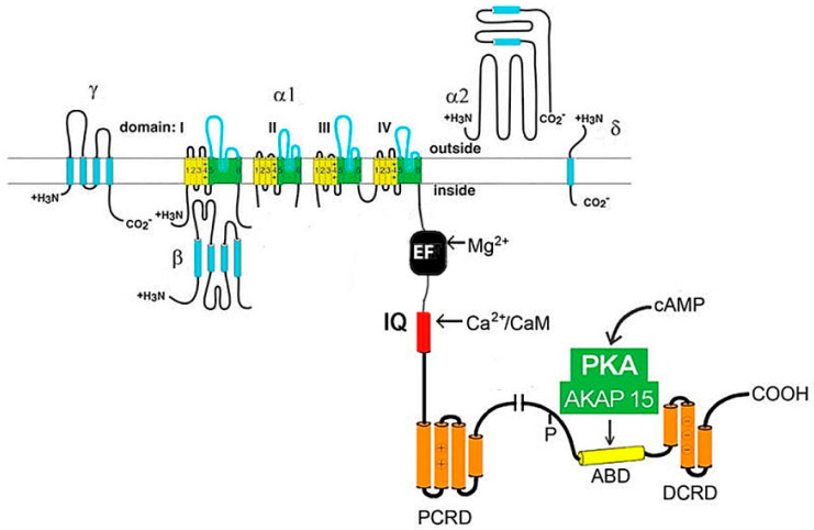 Fig. 1 Schematic topology of the CACNA1G-encoded CaV3.1 T-type calcium-channel α1G subunit, showing four homologous domains, voltage-sensing S4 segments and the Ca2+-permeable pore.1
The biological functions of transmembrane CACNA1G channel protein are focused on low-voltage activation, transient Ca2+ entry, and regulation of membrane excitability:
Creative Biolabs offers purified CACNA1G membrane samples produced under unified preparation workflows, including full-length CACNA1G constructs and isolated domain variants. Truncated domain fragments cannot support complete surface-partner-complex assembly behaviours, while full-length constructs suit research focused on channel-subunit-partner interaction and plasma-membrane anchoring functional observation. All batches receive uniform quality screening. Functional-relevant observation may only be carried out with full-length samples under simulated plasma-membrane microenvironment setups. Consistent structural features are preserved across batches to support comparative laboratory analysis across separate test groups. Full-length CACNA1G samples retain intact partner-interaction-domain conformation after standardized purification, which supports reliable detection of weak and transient subunit-partner contacts for comparative functional analysis.
Not finding the membrane protein product you need? Contact us to start your one-stop custom service!
Creative Biolabs provides adjustable CACNA1G expression cell research models with varied expression levels, applicable to structural observation of multi-pass transmembrane T-type low-voltage-activated calcium-channel proteins and research into membrane-partner molecular interaction. Sample evaluation includes sustained target expression detection and preliminary partner-interaction observation, enabling side-by-side comparison of subunit-binding behaviours under differing expression abundances. These cell systems can pair with diverse laboratory analysis schemes to track changes in partner-assembly efficiency alongside shifting target protein levels.
Not finding the stable cell line product you need? Contact us to start your one-stop custom service!
Anti-CACNA1G recombinant antibodies are generated via standardized protein preparation workflows, compatible with routine laboratory detection techniques for plasma-membrane localization mapping and identification of subunit-partner molecular complexes. The antibody series can work alongside common laboratory detection reagents to realize multi-dimensional observation of target distribution within membrane-enriched sample materials.
Not finding the recombinant antibody product you need? Contact us to start your one-stop custom service!
Beyond catalog products, Creative Biolabs offers specialized custom services for CACNA1G research:
CACNA1G might act as a multi-pass transmembrane T-type low-voltage-activated calcium-channel pore-forming α-subunit protein and participate in heteromeric plasma-membrane complex assembly to modulate membrane-channel-complex arrangement and plasma-membrane homeostasis.
CACNA1G expression status could alter membrane-partner-complex assembly efficiency and local plasma-membrane-coupled molecular-interaction balance, serving as a major channel mediator of membrane-ion-homeostasis-associated biological processes.
No, CACNA1G-associated research reagents from Creative Biolabs are exclusively built for exploring membrane-channel-dependent plasma-membrane ion-regulatory mechanisms, and shall not be deployed for any clinical-oriented workflows. These preparations are optimized for basic laboratory investigation and do not satisfy performance benchmarks required for clinical implementation.
Offerings include full-length CACNA1G membrane protein, target-specific recombinant antibodies and tunable expression cell research models, supporting research on plasma-membrane homeostasis and membrane-channel-mediated membrane-partner perception.
Laboratory observation schemes may include subunit-partner interaction related tests to analyse molecular-binding associated behaviors under simulated plasma-membrane environments.








