
Creative Biolabs is offering the most comprehensive services for antibody development projects. With strict regulation and effective execution, we are dedicated to providing the most valuable solutions to complete your projects.
HighlightsCreative Biolabs is offering the most comprehensive services for antibody development projects. With strict regulation and effective execution, we are dedicated to providing the most valuable solutions to complete your projects.
HighlightsCreative Biolabs has established a broad range of platforms for developing novel antibodies or equivalents. These cutting-edge technologies enable our scientists to meet your demands from different aspects and tailor the most appropriate solution that contributes to the success of your projects.
HighlightsWith deep understanding in antibody-related realms and extensive project experience, Creative Biolabs offers a variety of references to help you learn more about our capacities and achievements, including infographic, flyer, case study, peer-reviewed publications, and all kinds of knowledge that can assist your projects. You are also welcome to contact us directly for more specific solutions.
HighlightsGet a real taste of Creative Biolabs, one of the most professional custom service providers in the world. We are committed to providing highly customized comprehensive solutions with the best quality to advance your projects.
Highlights Loading...
All listed services and products are For Research Use Only. Do Not use in any diagnostic or therapeutic applications.
The CD7 gene is located in the 17q25.3 region of the human chromosome and encodes the T-cell antigen CD7 (CD7), also known as GP40 or Leu-9, a member of the immunoglobulin superfamily. CD7 is a single-pass type I transmembrane glycoprotein, with an extracellular region comprising an N-terminal immunoglobulin V-type domain followed by a proline-rich stalk that projects the Ig domain away from the membrane, a single transmembrane helix, and a short cytoplasmic tail containing a YEDM motif that mediates association with phosphatidylinositol 3-kinase (PI3K). Its molecular weight is approximately 40 kDa in the glycosylated form and it is predominantly expressed on the surface of thymocytes, mature T lymphocytes, and natural killer cells, with lower levels detected on pre-B lymphocytes, myeloid progenitors, and plasmacytoid dendritic cells. As a co-stimulatory receptor involved in T-cell activation, CD7 binds its ligand SECTM1 at the immune synapse, and upon engagement, the YEDM motif recruits PI3K, leading to the production of D-3 phosphoinositides, tyrosine phosphorylation of downstream signaling intermediates, and enhanced cytokine production including IL-2 and interferon-γ. CD7 also promotes integrin-mediated adhesion of T cells and NK cells to fibronectin and ICAM-1, facilitating cell-cell interactions during immune responses. The CD7 gene has multiple alleles documented in ClinVar, though its role as a disease-associated locus remains limited, with population data suggesting that heterozygous loss is well tolerated. The protein is palmitoylated at membrane-proximal cysteine residues. Therefore, CD7 is regarded as an important model for studying co-stimulatory receptor function and T-cell signaling regulation. Its functional analysis provides valuable tools for understanding the molecular mechanisms of immune synapse organization, PI3K-dependent signal transduction, and the regulatory networks governing lymphocyte activation.
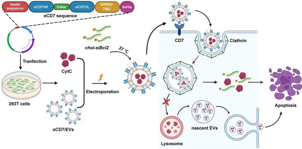 Fig. 1 Schematic diagram of the use of αCD7/EVs/CytC/siBcl2 for treating T-cell malignancies.1
The functional spectrum of CD7 centers on co-stimulatory signaling and immune synapse modulation in lymphocytes:
Creative Biolabs offers a comprehensive series of high-quality CD7 membrane protein products designed to support structural and functional investigations of co-stimulatory receptor signaling and immune synapse biology. Built upon the independently developed MemDX™ recombinant membrane protein technology platform, CD7 proteins can be prepared through multiple expression systems, including virus-like particles (VLP) and in vitro prokaryotic expression systems, among others. These rigorously characterized protein products, which include MemDX™ recombinant human CD7 membrane protein (as virus-like particles) and human CD7 proteins derived from diverse expression systems, are suitable for ELISA, antibody screening and identification, ligand binding experiments, and functional research, equipping investigators with dependable reagents for analyzing CD7-mediated co-stimulatory signaling and lymphocyte activation mechanisms.
Not finding the membrane protein product you need? Contact us to start your one-stop custom service!
Constructing a robust cell model is crucial for investigating CD7 function in T-cell co-stimulatory signaling. As a plasma membrane receptor that engages SECTM1 at the immune synapse and activates PI3K-dependent downstream pathways, the expression level and membrane distribution of CD7 directly affect the magnitude of co-stimulatory signals and the threshold for lymphocyte activation. Alterations in CD7 expression or changes in its immune synapse localization may lead to significant differences in T-cell responsiveness. Creative Biolabs offers customized CD7 membrane protein stable transfection cell models, covering wild-type and various functionally characterized forms. These models stably express the target protein on the plasma membrane, and are suitable for studies on SECTM1 ligand binding, immune synapse localization analysis, PI3K-AKT signaling pathway activation, and quantitative assessment of co-stimulatory signal output, providing a reliable and reproducible platform for exploring the molecular mechanism, structure-function relationship, and regulatory role of CD7 in lymphocyte activation. Our technology ensures correct protein localization and plasma membrane integrity, facilitating your research.
Not finding the stable cell line product you need? Contact us to start your one-stop custom service!
Creative Biolabs offers highly specific recombinant antibodies targeting CD7, produced using advanced recombinant antibody technology. Compared to traditional antibodies, these recombinant antibodies demonstrate superior performance in terms of specificity, sensitivity, and batch-to-batch consistency. They can also be modified and labeled in various ways. Our CD7 recombinant antibodies support a wide range of applications, including Western Blot, ELISA, flow cytometry, immunofluorescence, immunocytochemistry, immunohistochemistry, and immunoprecipitation, and can be used to detect differences in CD7 expression levels on various lymphocyte populations, analyze its plasma membrane distribution and immune synapse localization, and conduct related research such as ligand-receptor interaction analysis and PI3K signaling pathway investigation. These antibodies provide reliable detection tools for the structural analysis, functional study of CD7, and the investigation of its role in co-stimulatory signaling and T-cell immune regulation.
Not finding the recombinant antibody product you need? Contact us to start your one-stop custom service!
In addition to the standard products, Creative Biolabs also offers professional customized services for CD7. CD7 is a type I transmembrane glycoprotein with an extracellular Ig V-type domain and a proline-rich stalk that positions the ligand-binding site for optimal engagement at the immune synapse. These structural features present specific requirements in aspects such as membrane protein expression, antigen design, and functional characterization. Leveraging our technology platform, we can assist you in completing the following customized projects:
CD7 functions as a co-stimulatory receptor on the surface of T lymphocytes and natural killer cells, providing essential secondary signals during immune activation. Its extracellular Ig V-type domain binds the ligand SECTM1 at the immune synapse, while its cytoplasmic YEDM motif recruits and activates PI3K, triggering downstream signaling cascades that promote cytokine production, including IL-2 and interferon-γ, and enhance integrin-mediated cell adhesion. CD7 serves as an important model for studying co-stimulatory receptor biology and immune synapse signaling.
CD7 possesses a well-characterized signaling mechanism in which its YEDM motif directly couples to the PI3K-AKT pathway, providing a tractable system for dissecting co-stimulatory signal transduction. Investigating CD7 yields insights into immune synapse organization, the molecular logic of T-cell activation thresholds, and how co-stimulatory and inhibitory signals are integrated during lymphocyte responses. The receptor's restricted expression pattern on lymphoid cells and its role in modulating cytokine production and adhesion make it a valuable model for studying immune cell communication and regulation.
Not applicable. All CD7 products and services provided by Creative Biolabs are for research purposes only and cannot be used for any clinical diagnosis, prevention, treatment or cure of any disease.
We offer a variety of product types, including recombinant CD7 membrane proteins, stable cell models expressing CD7, high-quality recombinant CD7 antibodies, as well as comprehensive customized services.








