
Creative Biolabs is offering the most comprehensive services for antibody development projects. With strict regulation and effective execution, we are dedicated to providing the most valuable solutions to complete your projects.
HighlightsCreative Biolabs is offering the most comprehensive services for antibody development projects. With strict regulation and effective execution, we are dedicated to providing the most valuable solutions to complete your projects.
HighlightsCreative Biolabs has established a broad range of platforms for developing novel antibodies or equivalents. These cutting-edge technologies enable our scientists to meet your demands from different aspects and tailor the most appropriate solution that contributes to the success of your projects.
HighlightsWith deep understanding in antibody-related realms and extensive project experience, Creative Biolabs offers a variety of references to help you learn more about our capacities and achievements, including infographic, flyer, case study, peer-reviewed publications, and all kinds of knowledge that can assist your projects. You are also welcome to contact us directly for more specific solutions.
HighlightsGet a real taste of Creative Biolabs, one of the most professional custom service providers in the world. We are committed to providing highly customized comprehensive solutions with the best quality to advance your projects.
Highlights Loading...
All listed services and products are For Research Use Only. Do Not use in any diagnostic or therapeutic applications.
SIT1 is a small ~20–22 kDa type‐III single pass membrane well-characterized surface molecule found primarily expressed in hematopoietic lineages. SIT1 is composed of a short extracellular N-terminal region, one membrane-spanning alpha-helical domain and an extended cytoplasmic tail containing multiple immunoreceptor tyrosine-based inhibition motifs (ITIMs) as well as Src homology 2 (SH2) domain-binding sites. SIT1 is rapidly tyrosine phosphorylated upon engagement of the T-cell receptor (TCR) or B-cell receptor (BCR), generating docking sites for SHP-1 and -2 as well as Grb2, a mediator critical in regulating intracellular signalling events. This molecular assembly raises the qualitative threshold necessary for full lymphocyte activation by reducing proximal phosphorylation events, including ZAP-70, LAT, SLP-76 and PLCγ1; calcium mobilization downstream from TCR engagement as well as MAPK signaling and NF-κB nuclear translocation. In this capacity, SIT1 serves as an essential rheostat that inhibits spontaneous autoimmunity while maintaining the potential for strong pathogen-specific immune responses. Besides classical T and B lymphocytes, SIT1 regulates NK cell education, iNKTcell development and functional plasticity of tissue-resident memory lymphocytes. In clinical contexts, dysregulated expression of SIT1 or mutations at its phosphorylation sites have been implicated in systemic lupus erythematosus, rheumatoid arthritis and lymphoproliferative syndromes. Dysregulated SIT1 signaling in the tumor microenvironment may affect T-cell activation versus exhaustion balance, therefore impacting checkpoint inhibitor sensitivity. Together, these convergent immunological features position SIT1 as an essential target for biologics to restore tolerance and next-generation checkpoint modulation, but also precision autoimmune therapeutics.
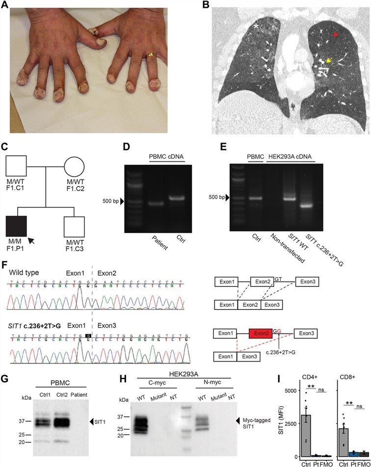 Fig.1 Identification of a patient with a SIT1 homozygous variant.1
SIT1 orchestrates multiple immune programs that go beyond its classical act as a simple signal attenuator:
We present a diverse panel of highly purified full-length and engineered SIT1 transmembrane protein constructs designed to support structural and functional studies of this immune adaptor. Because membrane topology, cytoplasmic tyrosine phosphorylation, and accessibility of signaling motifs can influence SIT1-mediated protein interactions, our protein production strategies are tailored to preserve key structural and biochemical properties relevant to downstream research. These SIT1 research constructs can support phosphorylation studies, protein–protein interaction assays, structural characterization, and screening applications focused on SIT1-associated signaling mechanisms. Protein format, preparation conditions, phosphorylation state, and applicable functional assays are selected and confirmed according to specific project requirements. Quality control and characterization strategies are likewise tailored to the intended experimental application.
Not finding the membrane protein product you need? Contact us to start your one-stop custom service!
Functionally relevant cell-based platforms that mimic the physiological cellular microenvironment are needed to decipher SIT1-mediated immunoregulatory thresholds and promote therapeutic pathways for enhanced adhesion. We provide the research community with well-established SIT1 stable cell lines characterized by authentic TCR/BCR plasma membrane localization along with status-neutral basal & inducible tyrosine phosphorylation capacity when either receptor is stimulated. Alongside wild-type SIT1, our platforms comprise constitutive ITIM mutants, disease-associated tyrosine to phenylalanine variants and reporter systems linking NFAT, NF-κB or IL-2 promoter elements with luciferase for real-time quantifying of signaling. We also offer knockdown rescue systems, and lines co-expressing SHP-1 or SHP-2 to interrogate the kinetics of phosphatase recruitment. All clones are stringently validated through verification of genomic integration, surface staining and functional validation in calcium flux, proliferation and T-cell activation assays. These scalable cellular resources provide critical substrates for studies of autoimmune mechanisms and screening compounds with immunomodulatory potential, as well as preclinical optimization of biologics antagonizing the SIT1 axis.
Not finding the stable cell line product you need? Contact us to start your one-stop custom service!
SIT1 monoclonal antibodies: enhance your SIT1 investigations with a focused selection of sequence-defined, high-affinity monoclonal antibodies developed to recognize SIT1. Depending on the individual clone, antibodies may recognize epitopes within different regions of SIT1 and support species-specific detection as documented in the corresponding datasheet. Validated applications may include Western blotting, ELISA, flow cytometric analysis, immunofluorescence and confocal microscopy, and immunohistochemistry, depending on the characteristics and validation data available for each antibody. These reagents can support studies of SIT1 expression across immune cell subsets, cellular localization, and signaling-associated changes. Epitope specificity, species reactivity, phosphorylation-state recognition, cross-reactivity, and suitability for functional assays should be determined according to the clone-specific datasheet and available experimental evidence.
Not finding the recombinant antibody product you need? Contact us to start your one-stop custom service!
Extending our standard catalog, we offer comprehensive, end-to-end design and engineering services purpose-built for SIT1-focused programs:
No—our SIT1 reagents including recombinant proteins, stable cell lines and antibody assays are produced solely for non-clinical research.
Yes, we have constructed stable lines in which SIT1 transcription is placed under a doxycycline-responsive promoter. This arrangement permits precise initiation of expression at defined time points and subsequent monitoring of progressive SHP-1 recruitment, CD3ζ chain dephosphorylation, and IL-2 transcriptional suppression with temporal resolution, eliminating the chronic T-cell exhaustion and compensatory PD-1 upregulation inherent to constitutive overexpression systems.
Yes. We will prepare SIT1 constructs for these purposes that are inserted as expected into phospholipid bilayers with the cytoplasmic tail in a position to stall recruiting of cellular phosphatases and/or immune receptor engagement.
Our products maintain both the tyrosine residues and membrane topology necessary for phosphorylated (inducible) SHP-1 / SHP-2 recruitment. Phospho-state profiling is also part of the quality control process to ensure these regulatory sites are intact and accessible.








