
Creative Biolabs is offering the most comprehensive services for antibody development projects. With strict regulation and effective execution, we are dedicated to providing the most valuable solutions to complete your projects.
HighlightsCreative Biolabs is offering the most comprehensive services for antibody development projects. With strict regulation and effective execution, we are dedicated to providing the most valuable solutions to complete your projects.
HighlightsCreative Biolabs has established a broad range of platforms for developing novel antibodies or equivalents. These cutting-edge technologies enable our scientists to meet your demands from different aspects and tailor the most appropriate solution that contributes to the success of your projects.
HighlightsWith deep understanding in antibody-related realms and extensive project experience, Creative Biolabs offers a variety of references to help you learn more about our capacities and achievements, including infographic, flyer, case study, peer-reviewed publications, and all kinds of knowledge that can assist your projects. You are also welcome to contact us directly for more specific solutions.
HighlightsGet a real taste of Creative Biolabs, one of the most professional custom service providers in the world. We are committed to providing highly customized comprehensive solutions with the best quality to advance your projects.
HighlightsFor over two decades, Creative Biolabs has been at the forefront of protein interaction analysis. As a trusted partner to the global scientific community, we offer a sophisticated suite of Protein-Fragment Complementation Assay (PCA) services, also known as split-protein complementation assays. Our platforms are engineered to provide robust, quantitative, and real-time analysis of protein-protein interactions (PPIs) directly within the complex milieu of living cells and even whole organisms. By harnessing the latest advancements in reporter technology, we empower researchers to unravel complex biological networks, validate drug targets, and screen for novel therapeutics with exceptional confidence.
The Protein-Fragment Complementation Assay is a powerful and versatile method for the direct detection of protein-protein interactions in vivo. The core principle is elegant in its simplicity: a reporter protein is rationally dissected into two non-functional fragments. These fragments are then genetically fused to two proteins of interest, "X" and "Y".
The reconstitution of the reporter generates a measurable signal (e.g., luminescence, fluorescence, or cell survival) that is directly proportional to the strength and abundance of the protein-protein interaction.
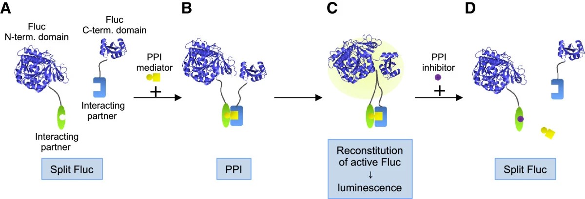 Fig. 1 Principle of Fluc PCA (A) Fluc is dissected into two parts, N-terminal domain and C-terminal domain.1
We offer a comprehensive selection of PCA systems, allowing us to select the optimal reporter for your specific cell type, protein localization, and experimental goals.
| PCA System | Principle | Signal Type | Key Advantages | Best For |
| Split-Luciferase | Interaction reconstitutes a luciferase enzyme. | Luminescence | Highest sensitivity, wide dynamic range, low background, ideal for quantitative measurements. | Detecting weak/transient interactions, HTS, in vivo imaging. |
| Bimolecular Fluorescence Complementation (BiFC) | Interaction reconstitutes a fluorescent protein (GFP, YFP, mCherry). | Fluorescence | Direct visualization of PPI localization via microscopy, stable signal. | Subcellular localization studies, confirming interactions in specific compartments. |
| Split-Ubiquitin System (SUS) | Interaction reconstitutes ubiquitin, leading to the cleavage and release of a transcription factor. | Reporter Gene (e.g., LacZ, HIS3) | Excellent for studying membrane protein interactions (Type I, II, multi-spanning). | GPCRs, transporters, ion channels, receptor-ligand interactions. |
| Dihydrofolate Reductase (DHFR) PCA | Interaction reconstitutes the DHFR enzyme, conferring resistance to methotrexate. | Cell Survival | Simple, selection-based readout; suitable for large-scale library screening. | Validating large sets of interactions, drug discovery screening. |
We have meticulously designed our PCA service workflow to be transparent, efficient, and collaborative.
Our PCA services are a versatile tool applicable to a wide array of research areas.
Our PCA services are not just an assay; they are a complete, end-to-end solution designed to deliver actionable insights.
PCA is a powerful tool, often best utilized as part of a multi-faceted research strategy. Creative Biolabs offers a comprehensive portfolio of services that complement our PCA platform. Explore our full range of PPI services here: Protein-Protein Interaction (PPI) Assay Services
Let's discuss how our advanced Protein-Fragment Complementation Assay services can accelerate your project.
Q: Which PCA system is best for my project?
A: The best system depends on your specific research question. For the highest sensitivity and quantitative results, we recommend split-luciferase. For visualizing interaction location, BiFC is ideal. For membrane proteins, the Split-Ubiquitin system is superior. Contact our specialists for a free consultation to determine the optimal approach.
Q: Can PCA be used to determine binding affinity (Kd)?
A: Yes, under carefully controlled conditions where protein expression levels are measured, dose-response curves can be generated to estimate the apparent affinity of the interaction within the cellular environment.
Q: What is the typical turnaround time for a PCA project?
A: Turnaround time varies based on project complexity, ranging from 4-6 weeks for simple pair validation to 8-12 weeks for a full high-throughput screening campaign.
Q: What do I receive in the final report?
A: You will receive a comprehensive report including the detailed experimental protocol, all raw and processed data, graphical representations of the results, statistical analysis, and a full scientific interpretation of the findings by our expert team.
All listed services and products are For Research Use Only. Do Not use in any diagnostic or therapeutic applications.








