
Creative Biolabs is offering the most comprehensive services for antibody development projects. With strict regulation and effective execution, we are dedicated to providing the most valuable solutions to complete your projects.
HighlightsCreative Biolabs is offering the most comprehensive services for antibody development projects. With strict regulation and effective execution, we are dedicated to providing the most valuable solutions to complete your projects.
HighlightsCreative Biolabs has established a broad range of platforms for developing novel antibodies or equivalents. These cutting-edge technologies enable our scientists to meet your demands from different aspects and tailor the most appropriate solution that contributes to the success of your projects.
HighlightsWith deep understanding in antibody-related realms and extensive project experience, Creative Biolabs offers a variety of references to help you learn more about our capacities and achievements, including infographic, flyer, case study, peer-reviewed publications, and all kinds of knowledge that can assist your projects. You are also welcome to contact us directly for more specific solutions.
HighlightsGet a real taste of Creative Biolabs, one of the most professional custom service providers in the world. We are committed to providing highly customized comprehensive solutions with the best quality to advance your projects.
HighlightsInvestigating caspase function in drug discovery is often hindered by challenges such as the energy-bias inherent in ATP-dependent reporters and the kinetic delays associated with traditional fluorogenic substrates. Creative Biolabs' In vitro Caspase Biochemical Assays address these limitations by offering a robust platform for profiling and screening inhibitors specifically targeting this crucial class of proteases. We provide high-resolution, real-time apoptotic data, enabling the rapid identification of potent lead compounds. This precision accelerates your drug discovery process, effectively bridging the critical gap between initial screening and successful clinical development.
Caspases serve as the central executioners of pathway-specific apoptosis, functioning as tightly regulated proteases that orchestrate the controlled dismantling of cellular components in response to intrinsic and extrinsic death signals. Precise biochemical characterization of caspase activity is therefore essential for elucidating apoptotic mechanisms, validating therapeutic candidates targeting cell death pathways, and identifying compounds that selectively modulate distinct caspase family members for oncology and beyond.
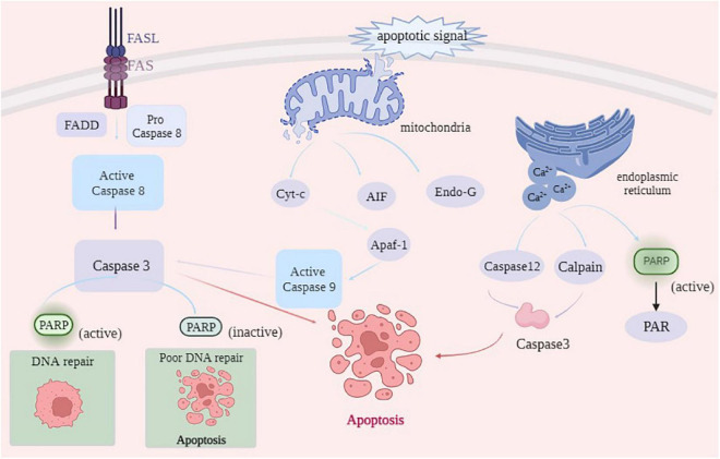 Fig.1 Core apoptotic pathways.1
Creative Biolabs provides a full panel of in vitro biochemical assays for precisely quantifying the activity of both initiator and executioner caspases. Moving beyond traditional endpoint measurements, our dynamic kinetic analysis captures the real-time "velocity" of apoptotic signaling. This approach enables more precise IC50 determinations and offers deeper mechanistic insights into drug-induced cytotoxicity.
Caspase-1 is a central mediator of inflammatory cell death (pyroptosis) and initiates innate immune responses by cleaving pro-IL-1β and pro-IL-18. Our assay employs a specific fluorogenic substrate in an enzyme-coupled reaction to quantify caspase-1 activity in cell lysates via real-time fluorescence measurement. With high sensitivity and specificity, this method effectively minimizes cross-reactivity with other caspases, making it ideal for evaluating inflammatory models and screening anti-inflammatory compounds.
As a highly conserved initiator caspase, caspase-2 plays a pivotal role in PIDDosome-mediated apoptosis induced by genotoxic stress. Our detection system utilizes a selective fluorogenic substrate that, upon cleavage by activated caspase-2, generates a quantifiable fluorescence signal. This technique sensitively captures early apoptotic events triggered by DNA damage or chemical modulators, offering particular value in mechanistic studies of genotoxic agents and small-molecule agonist screening.
Caspase-3 is the key executioner caspase in apoptosis, serving as the primary convergence point for both intrinsic and extrinsic pathways by cleaving essential cellular substrates. Our caspase-3 activity assay uses the well-established DEVD-AMC substrate, with fluorescence intensity monitored dynamically to reflect apoptotic progression. This robust, reproducible, and broadly applicable method supports a wide range of apoptotic models, including both adherent and suspension cells.
Caspase-4, an inflammatory caspase in humans, is central to the non-canonical inflammasome pathway, directly sensing intracellular lipopolysaccharide (LPS) and triggering pyroptosis with potential crosstalk to apoptosis. Our assay leverages its substrate specificity using LEVD-AMC for precise activity measurement. The key advantage lies in its ability to distinguish caspase-4 activation from downstream effector caspases-3/-7, providing a powerful tool for studying immune evasion in intracellular bacterial infections and validating anti-inflammatory targets.
Caspase-5 functions within the inflammasome network, cooperating with caspase-4 during bacterial infection or endogenous danger signaling to mediate pyroptosis and potentially engage apoptotic pathways. By optimizing buffer conditions and employing a specific fluorogenic substrate, our assay accurately captures caspase-5 activation dynamics even in complex biological samples. This approach is particularly suited for investigating the distinct roles of caspase-5 in inflammatory disease models and its regulatory interactions with intersecting pathways.
Caspase-6 is a unique effector caspase prominently activated in neurodegenerative disorders, where it cleaves tau and amyloid precursor proteins, contributing to axonal degeneration and neuronal apoptosis. Our activity assay capitalizes on its substrate specificity using highly sensitive fluorometric or chemiluminescent detection, enabling reliable quantification even in low-expression systems. This technology offers neuroscientists a precise tool to assess neuronal injury and evaluate the efficacy of candidate neuroprotective agents.
Caspase-7, a homolog of caspase-3, not only executes apoptosis but also specifically participates in cytoskeletal remodeling and inactivation of DNA repair proteins. Our optimized DEVDase assay incorporates a selective caspase-7 inhibitor control, allowing precise discrimination of caspase-7's contribution from total DEVD-cleaving activity. This refined approach is especially valuable for detailed apoptotic pathway dissection and studies distinguishing caspase-3 and caspase-7 functions.
Caspase-8 is the initiating switch of the extrinsic apoptotic pathway, recruited to the DISC upon death receptor (e.g., Fas) activation to trigger cascade signaling, while its activity also critically suppresses necroptosis. Our caspase-8 activity assay employs the IETD-AMC substrate alongside a proprietary reaction buffer to maintain enzymatic stability. The core strength of this service lies in its ability to assess apoptotic sensitivity induced by death ligands (e.g., TRAIL) and elucidate the molecular switch mechanisms governing cell survival and death decisions.
Caspase-9 is the essential initiator of the mitochondria-mediated intrinsic apoptotic pathway, activated within the apoptosome to trigger downstream effector caspase cascades. Our service utilizes fluorogenic substrate technology for specific detection of caspase-9 activation following apoptosome formation. With an exceptionally high signal-to-noise ratio, this assay effectively reflects mitochondrial stress, cytochrome c release, and regulatory effects of anti-apoptotic proteins such as cIAP1, making it ideal for evaluating drug-induced mitochondrial toxicity and investigating anticancer mechanisms.
Caspase-10 cooperates with caspase-8 in the human extrinsic apoptotic pathway, recruited to the death-inducing signaling complex (DISC) to initiate apoptosis and possessing the ability to cleave and activate downstream effector caspases. Our caspase-10 activity assay employs a specific fluorogenic substrate with continuous kinetic readout to quantify its activation under apoptosis-inducing conditions. This methodological advantage enables investigation of apoptotic defects in Fas/TRAIL-resistant tumor cell lines and supports evaluation of novel anticancer agents targeting DISC components.
With over 20 years of expertise, our advanced platform eliminates traditional assay interference. Benefit from ATP-independent reporters for reliable post-apoptotic data, a superior 10-fold signal-to-noise ratio minimizing false positives, and HTS-validated assays ready for massive compound library screening.
Unlock the full potential of your apoptosis research with our advanced, ATP-independent assays. Contact our expert team now to discuss your specific project needs and discover how our HTS-ready platforms can accelerate your drug discovery timeline. Email us today for a personalized consultation and quote.
Reference
All listed services and products are For Research Use Only. Do Not use in any diagnostic or therapeutic applications.








