
Creative Biolabs is offering the most comprehensive services for antibody development projects. With strict regulation and effective execution, we are dedicated to providing the most valuable solutions to complete your projects.
HighlightsCreative Biolabs is offering the most comprehensive services for antibody development projects. With strict regulation and effective execution, we are dedicated to providing the most valuable solutions to complete your projects.
HighlightsCreative Biolabs has established a broad range of platforms for developing novel antibodies or equivalents. These cutting-edge technologies enable our scientists to meet your demands from different aspects and tailor the most appropriate solution that contributes to the success of your projects.
HighlightsWith deep understanding in antibody-related realms and extensive project experience, Creative Biolabs offers a variety of references to help you learn more about our capacities and achievements, including infographic, flyer, case study, peer-reviewed publications, and all kinds of knowledge that can assist your projects. You are also welcome to contact us directly for more specific solutions.
HighlightsGet a real taste of Creative Biolabs, one of the most professional custom service providers in the world. We are committed to providing highly customized comprehensive solutions with the best quality to advance your projects.
HighlightsWhat is In Vivo Phage Display?
The in vivo phage display method applies bacteriophages to identify and isolate proteins or peptides which bind tightly to a target molecule inside living organisms. Phage-displayed peptide or protein libraries are screened within living organisms like mice, rats or humans during in vivo phage display as opposed to in vitro phage display which occurs outside living systems.
This technique uses bacteriophage to help select peptides or proteins with strong binding affinities within physiological conditions to discover new targets for drug development and diagnostic tool development.
Importance in Biotechnology and Drug Discovery
Phage display performed in living organisms shows great potential for advancing biotechnology through its applications in drug discovery and development. The approach enables researchers to isolate new biomolecular interactions in living organisms and provides clear benefits compared to traditional antibody production methods and random peptide libraries. The technique stands out for identifying therapeutic peptides and antibodies able to bind disease-specific targets which researchers can subsequently develop into cancer treatment drugs and other autoimmune disease medications.
In vivo phage display helps researchers bypass key limitations of in vitro methods that include inadequate physiological environment simulation and limited target receptor expression in cell cultures. In vivo phage display technology generates precise models of molecular activity within organisms and improves therapeutic outcomes through targeted treatment approaches.
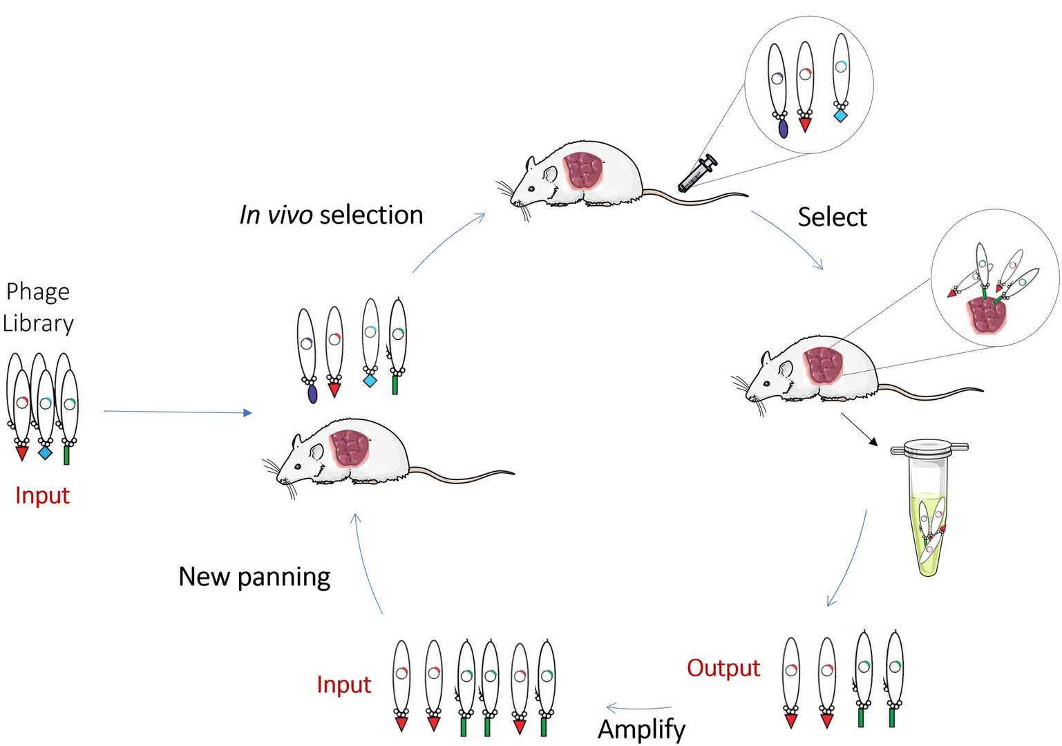 Fig. 1 Schematic representation of in vivo Phage Display.1, 3
Basic Principles of Phage Display
Phage display works through the genetic alteration of bacteriophages to make foreign peptides and proteins display on their surfaces on their surfaces. Researchers genetically link these peptides or proteins to either surface protein pIII or pVIII of the bacteriophage. By examining an extensive library of peptides displayed on the phage surface, researchers can discover peptides that bind to their specific target of interest.
The basic phage display process consists of several key steps.
How In Vivo Phage Display Works
In vivo phage display extends traditional phage display techniques by performing the procedure inside living organisms like mice or rats. The initial step of the technique involves injecting animals with a diverse collection of phages through either intravenous or intraperitoneal routes. These phages circulate through the blood system and bind to particular target molecules inside the body including cell receptors, tumor antigens and proteins that become overexpressed during disease conditions.
Researchers can extract phages from tissues, organs or blood following their binding to specific targets. Scientists perform isolation of the phages followed by sequencing of their genetic material which codes for the peptides or proteins responsible for target binding. Scientists obtain a collection of potential peptides and proteins suitable for subsequent therapeutic and diagnostic research.
 Fig. 2 In vivo phage display using differential binding approach.2, 3
In Vivo Phage Display Library Screening
During in vivo phage display screening researchers inject phage libraries into living organisms like mice to isolate phages that attach to specific target sites within tissues or organs. The targets identified through this process may involve proteins, peptides or cell surface receptors that play a role in diseases like cancer or inflammation.
Key Steps in Phage Library Screening:
 Fig. 3 Flow of in vivo phage biopanning.2, 3
In Vivo Phage Display Selection Process
The selection method works to increase the number of phages which bind exclusively to targeted molecular markers. Multiple rounds of binding, washing and amplification during in vivo selection produce a final pool consisting exclusively of high-affinity binders.
Methodologies for In Vivo Phage Display
Different optimization techniques exist to improve the effectiveness of in vivo phage display. Some of the common methods include:
Phage Display Platforms: Overview of Available Systems
The collection of phage display libraries presents multiple formats such as:
The choice of which library to use should be determined by the specific application needs, such as therapeutic discovery or diagnostic purposes and basic research investigations.
Therapeutic Applications: Targeted Drug Delivery
Targeted drug delivery systems have been effectively developed using in vivo phage display. Researchers who choose phages that attach to disease-specific biomarkers or receptors can guide therapeutic agents such as chemotherapy drugs or siRNAs to targeted cells which reduces unintended effects and strengthens treatment effectiveness.
Vaccine Development
Vaccine development requires extensive utilization of phage display technology. Through the identification of immune-stimulating peptides or proteins within phage display libraries, researchers can develop vaccines against infectious diseases and cancer. Peptides displayed on phages serve as potential antigen candidates when researchers develop vaccines that aim to prevent and treat diseases.
Diagnostic Applications
Diagnostic assays benefit from in vivo phage display through the identification of disease-specific biomarkers. Diagnostic kits use phages that attach to biomarkers to identify diseases including cancer and infections.
Biochemical Research and Screening
The technique of in vivo phage display allows scientists to study how proteins and peptides interact within living organisms. This technology reveals new binding interactions which help researchers understand cellular signaling pathways together with enzyme functions and receptor-ligand interactions.
Pros and Cons of In Vivo Phage Display and In Vitro Phage Display
Phage display technology, a powerful tool in biotechnology and drug discovery, can be performed in two main formats: in vivo and in vitro. The fundamental principle of displaying peptides, proteins or antibodies on bacteriophages remains the same for both approaches but their selection processes occur in different environments. In the following section we will conduct a detailed comparison between the two methods to evaluate their strengths and weaknesses as well as determine the ideal contexts for their application.
Table 1. Comparison of Pros and Cons of in vivo phage display versus in vitro phage display.
| Aspect | In Vivo Phage Display | In Vitro Phage Display |
| Physiological Relevance | High accuracy, mimics real biological environments. | Lacks true physiological context and immune responses. |
| Identification of Functional Binders | Enables discovery of peptides that exhibit functional binding. | Limited to identifying binders that work in isolated systems. |
| Better Targeting | Can target specific tissues, e.g., tumors or inflammatory sites. | Cannot target specific tissues effectively due to lack of biological mimicry. |
| Longer Half-life and Stability | Phages are more stable and have longer half-life. | Phages may not exhibit real-life stability in vitro. |
| Ethical Concerns | Ethical concerns about animal use, and welfare considerations. | No ethical concerns, as no animal models are involved. |
| Higher Complexity and Cost | More complex and expensive; requires animal models and specialized equipment. | Simpler, less expensive; requires no animal handling or specialized equipment. |
| Limited Throughput | Slower and more time-consuming due to multiple selection rounds. | Faster and more scalable, with potential for high-throughput screening. |
| Risk of Off-Target Effects | Potential for non-specific binding and false positives due to complex environment. | Less risk of non-specific binding compared to in vivo, but still possible. |
| Speed and Scalability | Slower compared to in vitro, requiring animal handling and monitoring. | Fast and efficient with the ability to screen large libraries quickly. |
| Lower Cost | Higher cost due to animal use, monitoring, and imaging. | Lower cost due to no need for animal models or complex equipment. |
| Simplified Experimental Setup | Requires handling living organisms, which can be unpredictable. | Easier to set up and control experimental conditions. |
| Broader Range of Targets | Can lead to false positives due to the lack of in vivo interaction. | Can be used to target isolated molecules or proteins on surfaces. |
| Lack of Physiological Context | Requires the biological complexity that may not be represented in vitro. | Misses the physiological context of how molecules interact in living systems. |
| No Immune or Metabolic Response | Lack of immune and metabolic processing limits discovery of functional binders. | Does not account for immune responses or metabolic processes. |
| Limited Discovery of Functional Binders | Not as effective at identifying functional interactions without physiological context. | Fails to identify peptides or proteins with functional effects in vivo. |
| Inability to Mimic Targeted Delivery | Challenges in mimicking real-world delivery systems like blood-brain barrier. | Does not simulate how molecules behave in real-world conditions like delivery barriers. |
Case Studies: In Vivo vs. In Vitro Phage Display Success Stories
Case Study 1: The in vivo phage display technique successfully identified peptides that bind tumor vasculature through a specific study. Through in vivo screening in mouse models scientists discovered peptides which exhibited high specificity for tumor sites to enable the delivery of chemotherapeutic agents to these areas. The in vitro phage display approach demonstrated lower effectiveness in identifying these peptides because it failed to replicate the tumor microenvironment and vasculature conditions accurately.
Case Study 2: Researchers used in vivo phage display to identify human antibodies that attach to a tumor antigen in another study on antibody discovery for cancer immunotherapy. Animal models were used to evaluate these antibodies for tumor growth inhibition ability. The in vitro phage display method successfully identified antibodies but failed to reveal their effectiveness at targeting cancer cells in living organisms thus demonstrating the critical role of biological context in therapeutic antibody research.
All listed services and products are For Research Use Only. Do Not use in any diagnostic or therapeutic applications.








