
Creative Biolabs is offering the most comprehensive services for antibody development projects. With strict regulation and effective execution, we are dedicated to providing the most valuable solutions to complete your projects.
HighlightsCreative Biolabs is offering the most comprehensive services for antibody development projects. With strict regulation and effective execution, we are dedicated to providing the most valuable solutions to complete your projects.
HighlightsCreative Biolabs has established a broad range of platforms for developing novel antibodies or equivalents. These cutting-edge technologies enable our scientists to meet your demands from different aspects and tailor the most appropriate solution that contributes to the success of your projects.
HighlightsWith deep understanding in antibody-related realms and extensive project experience, Creative Biolabs offers a variety of references to help you learn more about our capacities and achievements, including infographic, flyer, case study, peer-reviewed publications, and all kinds of knowledge that can assist your projects. You are also welcome to contact us directly for more specific solutions.
HighlightsGet a real taste of Creative Biolabs, one of the most professional custom service providers in the world. We are committed to providing highly customized comprehensive solutions with the best quality to advance your projects.
HighlightsModern biotherapeutics, diagnostics, and research tools all depend heavily upon antibodies as fundamental components. The unique specificity and effective functions of antibodies originate from their primary amino acid sequences because these sequences determine their three-dimensional formation as well as their antigen-binding properties and stability while also influencing their immunogenic potential. During antibody discovery and development absolute accuracy of amino acid sequences is essential. This precise sequence information is critical for:
De novo antibody sequencing, the process of determining the sequence without prior genetic information, is indispensable when the original hybridoma cell line is lost or unavailable, or when characterizing antibodies from complex biological samples.
Mass spectrometry has advanced protein analysis yet remains unable to differentiate between isobaric amino acids which share identical nominal and exact mass values. Isoleucine (Ile, I) paired with Leucine (Leu, L) stands out as the most notorious and commonly encountered pair during protein sequencing. Both molecules share the chemical composition C6H13NO2 and feature a monoisotopic mass of roughly 113.08406 Da. MS/MS employs standard Collision-Induced Dissociation (CID) or Higher-energy Collisional Dissociation (HCD) fragmentation techniques which target the peptide backbone to generate b- and y-ions. Peptides with substitutions between Ile and Leu create CID/HCD fragment ions with identical masses which prevents their definitive identification using these methods.
Despite their identical mass, Ile and Leu are structural isomers with distinct side chain arrangements:
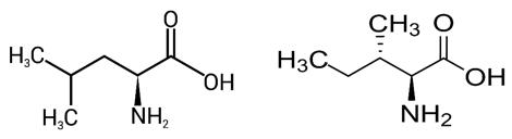 Fig. 1 Structure of Leucine and Isoleucine.
This subtle difference in side chain topology can have significant functional consequences:
The hydrophobic amino acids Ile and Leu occur frequently within protein structures and antibody variable domains. These amino acids consistently show up within the six CDR loops (H1, H2, H3, L1, L2, L3), which create the main antigen-binding surface. The specific sequence and structure of these loops dictate antigen specificity.
| Amino Acid | Typical Frequency Range in CDRs | Notes |
| Serine (S) | High | Often involved in H-bonding |
| Glycine (G) | High | Provides flexibility |
| Tyrosine (Y) | Moderate-High | Bulky, aromatic, H-bonding potential |
| Aspartate (D) | Moderate | Charged, H-bonding |
| Leucine (L) | Moderate-High | Hydrophobic core/contacts |
| Isoleucine (I) | Moderate | Hydrophobic core/contacts, β-branched |
| Arginine (R) | Moderate | Charged, H-bonding |
| Alanine (A) | Moderate | Small, hydrophobic |
| ... | ... | ... |
Given their prevalence in these functionally critical regions, accurately identifying each Ile and Leu position is non-negotiable for understanding and engineering antibody binding.
Failing to correctly distinguish between Ile and Leu during de novo sequencing can lead to severe downstream consequences:
Resolving the Ile/Leu ambiguity requires specialized techniques beyond standard Collision-Induced Dissociation (CID) or Higher-energy Collisional Dissociation (HCD) fragmentation. At Creative Biolabs, we employ a multi-pronged strategy integrating advanced MS methods, enzymatic digestion strategies, and sophisticated data analysis.
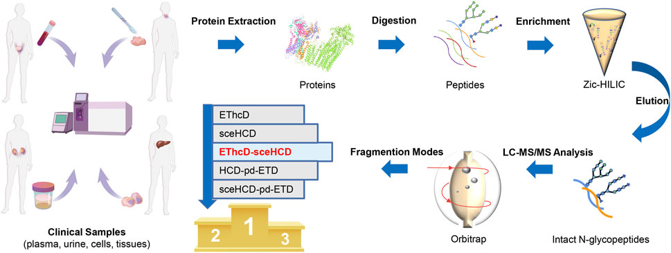 Fig. 2 Schematic illustration of the workflow for human plasma, urine, cells, and tissue intact N-glycopeptides analysis using different dissociation methods.1
The key to MS-based differentiation lies in fragmentation techniques that induce cleavage within the amino acid side chain, generating fragment ions whose masses are dependent on the specific Ile/Leu structure, unlike standard CID/HCD which primarily cleave the peptide backbone yielding b- and y-ions insufficient for this distinction.
Electron Transfer Dissociation (ETD) and Electron Transfer High Energy Collision Dissociation (EThcD) are powerful techniques in this context. Unlike CID/HCD which energize the entire peptide, ETD utilizes radical anions to transfer an electron to the protonated peptide precursor. This induces fragmentation predominantly at the peptide backbone N-Cα bond, generating c- and z•-type fragment ions. Crucially, ETD is a non-ergodic process, meaning fragmentation occurs faster than energy randomization, often preserving labile structures like post-translational modifications and, importantly, intact amino acid side chains on the resulting fragment ions. EThcD complements this by combining ETD activation with supplemental HCD activation, promoting more extensive fragmentation and potentially generating both b/y and c/z ions, along with side-chain specific fragments, providing richer data.
The true power of ETD/EThcD for Ile/Leu differentiation comes from the ability to induce secondary fragmentation of the radical z• ions (or through other pathways). This secondary fragmentation can cleave the Cα-Cβ bond or bonds within the side chain itself. This process generates specific neutral losses from the side chain, resulting in ions sometimes referred to as w-ions (though nomenclature can vary). The masses of these w-ions (or the mass differences corresponding to the neutral losses) are diagnostic for Ile vs. Leu:
Observing a fragment ion corresponding to a z-ion minus ~29 Da strongly suggests Isoleucine, while observing a z-ion minus ~43 Da points definitively to Leucine.
To enhance the generation and detection of these diagnostic side-chain fragments, Multi-stage Mass Spectrometry (MSn, typically MS³) can be employed. In an MS³ experiment, a specific information-rich fragment ion from the initial MS/MS (e.g., a z• ion containing the ambiguous Ile/Leu position) is isolated and subjected to further fragmentation (often CID or HCD). This process helps to generate the diagnostic w-ions with greater specificity and reduced background noise.
While MS provides the most direct evidence, enzymatic digestion strategies offer complementary information. Some proteases exhibit subtle preferences or hindered cleavage around branched aliphatic residues. For instance:
Observing consistently lower yields of a specific peptide bond cleavage across multiple experiments when using an enzyme like Chymotrypsin might suggest the presence of Ile rather than Leu at the P1 position, but this is rarely definitive on its own and serves primarily as supporting evidence.
A cornerstone of robust de novo sequencing is the use of multiple proteases with different specificities (e.g., Trypsin, Chymotrypsin, GluC, AspN, Elastase, Pepsin). This strategy generates a diverse pool of overlapping peptides. The benefits are twofold:
At Creative Biolabs, we recognize that accurate Ile/Leu determination is non-negotiable for reliable antibody sequencing. Our proprietary de novo sequencing platform, often incorporating approaches like our Database Assisted Shotgun Sequencing (DASS) methodology, is built upon a foundation designed to tackle this specific challenge. Creative Biolabs delivers antibody sequences with unmatched precision while definitively distinguishing between Isoleucine and Leucine to meet our clients' research and development demands.
Learn more about Creative Biolabs' de novo antibody sequencing services:
All listed services and products are For Research Use Only. Do Not use in any diagnostic or therapeutic applications.








