
Creative Biolabs is offering the most comprehensive services for antibody development projects. With strict regulation and effective execution, we are dedicated to providing the most valuable solutions to complete your projects.
HighlightsCreative Biolabs is offering the most comprehensive services for antibody development projects. With strict regulation and effective execution, we are dedicated to providing the most valuable solutions to complete your projects.
HighlightsCreative Biolabs has established a broad range of platforms for developing novel antibodies or equivalents. These cutting-edge technologies enable our scientists to meet your demands from different aspects and tailor the most appropriate solution that contributes to the success of your projects.
HighlightsWith deep understanding in antibody-related realms and extensive project experience, Creative Biolabs offers a variety of references to help you learn more about our capacities and achievements, including infographic, flyer, case study, peer-reviewed publications, and all kinds of knowledge that can assist your projects. You are also welcome to contact us directly for more specific solutions.
HighlightsGet a real taste of Creative Biolabs, one of the most professional custom service providers in the world. We are committed to providing highly customized comprehensive solutions with the best quality to advance your projects.
HighlightsPeptides and proteins serve as essential components in molecular biology while carrying out numerous vital functions necessary for life. Deciphering their structure stands as a crucial step because the linear amino acid sequence provides the basic knowledge layer.
Amino acids are the building blocks. Connecting amino acids through peptide bonds creates chains.
Size differences and structural complexity primarily distinguish these molecules. The peptide bond represents the primary connection between molecules while the amino acid sequence serves as their defining feature.
An amide linkage known as the peptide bond develops between the carboxyl group (-COOH) of one amino acid and the amino group (-NH₂) of the subsequent amino acid. During this condensation reaction water is released as a byproduct.
The backbone structure (-N-Cα-C-) maintains planarity and rigidity from its partial double bond nature which affects polypeptide folding patterns. Peptide sequencing aims to discover the bonds between amino acids and determine the order of amino acids along the chain.
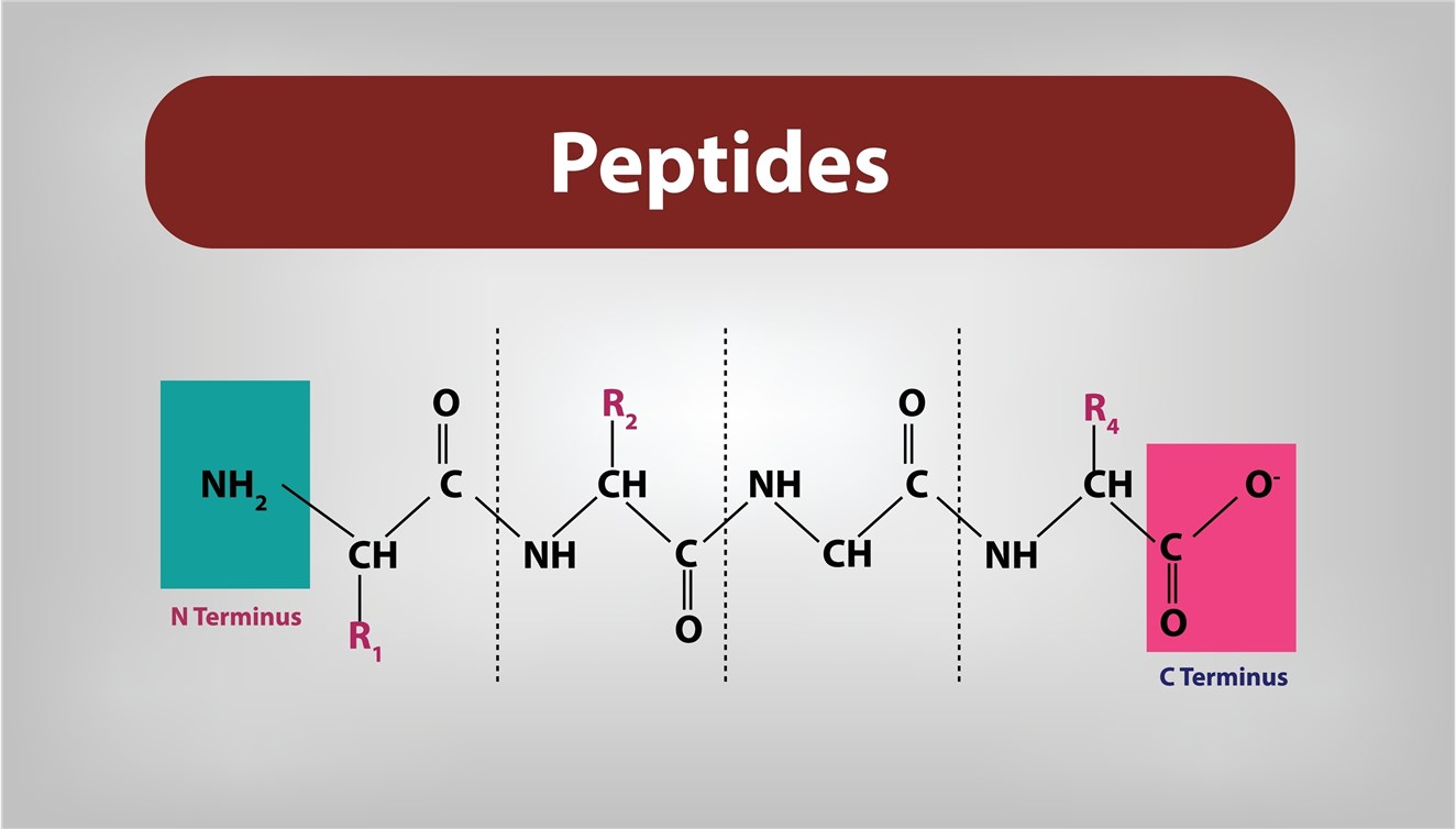 Fig. 1 Peptide structure.
The primary structure dictates:
A deeper dive into the components and structure is essential before exploring sequencing methodologies.
Twenty standard amino acids are encoded by the genetic code, each possessing a central carbon atom bonded to:
The R-group determines the amino acid's specific chemical properties (e.g., size, charge, hydrophobicity, reactivity), influencing the peptide's overall characteristics.
As mentioned, peptide bond formation is a dehydration synthesis reaction catalyzed by ribosomes during protein biosynthesis. In synthetic peptide chemistry, chemical coupling agents facilitate this process.
Chains are directional, possessing:
By convention, peptide sequences are written from the N-terminus to the C-terminus.
This linear arrangement (e.g., Ala-Gly-Ser-Met...) is the primary structure. Determining this exact order is the objective of peptide sequencing.
Developed by Pehr Edman in the 1950s, Edman degradation was the cornerstone of protein sequencing for decades. It provides direct sequence information from the N-terminus.
Edman degradation is a cyclical chemical process:
Each of the 20 standard amino acids yields a unique PTH derivative with a characteristic retention time on a specific HPLC column. By running known PTH-amino acid standards, a chromatogram library is created. The PTH-amino acid released in each cycle is identified by matching its peak's retention time to these standards.
| Feature | Strengths | Limitations |
| Methodology | Direct sequencing from N-terminus | Sequential, relatively slow (typically ~1 hour per cycle) |
| Sample Req. | Can work with relatively pure samples | Requires purified peptide/protein; μg to mg quantities often needed |
| Peptide Length | Reliable for ~30-50 residues per run | Incomplete reactions/side reactions accumulate, limiting read length |
| N-Terminus | Excellent for confirming N-terminal sequence | Fails if N-terminus is chemically blocked (e.g., acetylation, pyroglutamate) |
| Modifications | Poorly suited for identifying most PTMs | Most modifications interfere with the chemistry or identification |
| Mixtures | Generally unsuitable for complex mixtures | Requires single, pure sequence for clear results |
| Automation | Automated sequencers are available | Still requires significant hands-on time for setup and analysis |
While powerful, Edman degradation has been largely superseded by mass spectrometry for high-throughput and complex analyses, though it remains valuable for specific applications like N-terminal confirmation of recombinant proteins.
Mass spectrometry has revolutionized proteomics and peptide sequencing, offering unparalleled sensitivity, speed, and the ability to analyze complex mixtures and post-translationally modified peptides.
A mass spectrometer measures the mass-to-charge ratio (m/z) of ions. The basic workflow involves:
This is the workhorse of MS-based sequencing.
Fragmentation typically produces b-ions (containing the N-terminus) and y-ions (containing the C-terminus). The mass difference between consecutive ions in a b-series or y-series corresponds to the mass of a specific amino acid residue. By analyzing the pattern of fragment ions (the MS/MS spectrum), the amino acid sequence can be deduced.
Complex samples such as tryptic digests of whole proteomes undergo initial peptide separation through liquid chromatography, particularly Reversed-Phase HPLC (RP-HPLC), before mass spectrometric analysis.
LC-MS/MS technology enables researchers to identify and sequence numerous peptides from complicated mixtures through one experiment.
De novo sequencing directly identifies peptide sequences from MS/MS spectra without the need for a reference sequence database.
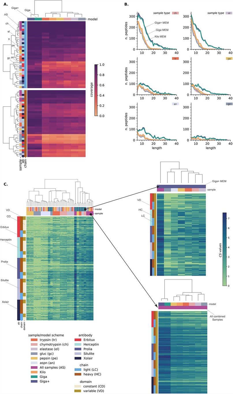 Fig. 2 Complete de novo sequencing of commercial monoclonal antibodies by deep learning models.1
While MS/MS fragmentation is effective, achieving complete fragmentation across every peptide bond (full b- and y-ion series) is rare. Gaps in the series can make definitive sequencing difficult, especially for de novo efforts. Proline residues, due to their cyclic structure, can lead to unique fragmentation patterns or suppress fragmentation altogether.
Peptide and protein amino acid sequencing serves as a fundamental element in both biological research and biopharmaceutical development today. Creative Biolabs utilizes decades of experience with state-of-the-art equipment and advanced LC-MS/MS systems to produce precise peptide sequencing data. Our commitment enables us to deliver the essential sequence information required by our clients to speed up their research breakthroughs. Meanwhile, we offer de novo antibody sequencing and de novo protein sequencing services, powered by our propriety DASS (Database Assisted Shotgun Sequencing) technology to meet the diverse protein research needs of our clients, driving innovation and advancement in the field of biomedical science.
Learn more about Creative Biolabs' de novo antibody sequencing services:
All listed services and products are For Research Use Only. Do Not use in any diagnostic or therapeutic applications.








