
Creative Biolabs is offering the most comprehensive services for antibody development projects. With strict regulation and effective execution, we are dedicated to providing the most valuable solutions to complete your projects.
HighlightsCreative Biolabs is offering the most comprehensive services for antibody development projects. With strict regulation and effective execution, we are dedicated to providing the most valuable solutions to complete your projects.
HighlightsCreative Biolabs has established a broad range of platforms for developing novel antibodies or equivalents. These cutting-edge technologies enable our scientists to meet your demands from different aspects and tailor the most appropriate solution that contributes to the success of your projects.
HighlightsWith deep understanding in antibody-related realms and extensive project experience, Creative Biolabs offers a variety of references to help you learn more about our capacities and achievements, including infographic, flyer, case study, peer-reviewed publications, and all kinds of knowledge that can assist your projects. You are also welcome to contact us directly for more specific solutions.
HighlightsGet a real taste of Creative Biolabs, one of the most professional custom service providers in the world. We are committed to providing highly customized comprehensive solutions with the best quality to advance your projects.
HighlightsSanger's protein sequencing method, developed by British biochemist Frederick Sanger and his colleagues in the late 1940s and early 1950s, is a chemical technique designed to determine the linear sequence of amino acids in a polypeptide chain. The core principle involves selectively labeling the N-terminal amino acid of a protein or peptide, followed by the complete hydrolysis of the polypeptide into its constituent amino acids. The labeled N-terminal amino acid is then identified, typically by chromatography. While this method identifies only one amino acid per cycle (the N-terminal one), through iterative application on overlapping peptide fragments, the entire sequence of a protein could be painstakingly reconstructed.
Prior to Sanger's work, the precise chemical nature of proteins was a subject of considerable debate. While it was known that proteins were composed of amino acids, the idea that each protein had a unique, genetically determined, and invariant sequence of these amino acids was not universally accepted. Some theories proposed proteins might be complex colloidal aggregates with no fixed structure.
Sanger's successful sequencing of insulin, completed in 1955, was a landmark achievement for several reasons:
The Sanger method, while laborious by modern standards, was elegant in its chemical logic for identifying the N-terminal amino acid of a polypeptide.
The fundamental principle relies on the chemical derivatization of the free α-amino group present at the N-terminus of a polypeptide chain. This derivatization creates a stable, identifiable tag on the first amino acid. Subsequent hydrolysis breaks all peptide bonds, releasing all amino acids, but the N-terminal one remains tagged and can be isolated and identified.
The key reagent employed by Sanger was 1-fluoro-2,4-dinitrobenzene, often referred to as 2,4-dinitrofluorobenzene (DNFB) or Sanger's reagent.
DNFB is highly reactive towards nucleophilic groups, particularly primary and secondary amines, under mild conditions. The fluorine atom is a good leaving group, facilitating nucleophilic aromatic substitution.
The polypeptide is reacted with DNFB, typically under mildly alkaline conditions (e.g., sodium bicarbonate solution, pH ~8-9). At this pH, the N-terminal α-amino group is largely deprotonated and thus highly nucleophilic. It attacks the carbon atom to which the fluorine is attached in DNFB, displacing the fluoride ion and forming a stable 2,4-dinitrophenyl (DNP) derivative of the N-terminal amino acid. This DNP-peptide is typically yellow due to the dinitrophenyl group.
Other free amino groups in the polypeptide, such as the ε-amino group of lysine side chains or a free N-terminus of another chain if the protein is multimeric, will also react with DNFB. However, only the α-amino group of the N-terminal amino acid will yield a DNP-amino acid that was originally at the N-terminus of that specific chain.
After the labeling reaction, the DNP-polypeptide is subjected to complete acid hydrolysis. This is typically achieved by heating the DNP-polypeptide in a strong acid solution (e.g., 6 M HCl at 100-110°C for several hours, typically 12-24 hours). This treatment cleaves all the peptide bonds within the polypeptide chain, releasing the individual amino acids.
The DNP group forms a very stable covalent bond with the N-terminal amino acid's α-nitrogen, a bond that is resistant to acid hydrolysis. Therefore, after hydrolysis, the N-terminal amino acid is recovered as its DNP-derivative (DNP-amino acid), while all other amino acids are released in their free, unmodified form.
The mixture resulting from hydrolysis contains free amino acids and the yellow DNP-amino acid (or DNP-derivatives of certain side chains like lysine, if present). The DNP-amino acid is then separated from the free amino acids and identified.
The successful application of this method to determine the complete amino acid sequence of bovine insulin was a monumental task that took Sanger and his team nearly a decade (from roughly 1945 to 1955).
Bovine insulin is a relatively small protein, but it presented complexities. It consists of two distinct polypeptide chains:
These two chains are covalently linked by two inter-chain disulfide bonds. The A-chain also contains one intra-chain disulfide bond.
Sanger's approach to sequencing insulin was systematic and meticulous:
The most significant limitation of the Sanger method was its destructive nature. In each cycle of labeling and hydrolysis:
Only the N-terminal amino acid was identified.
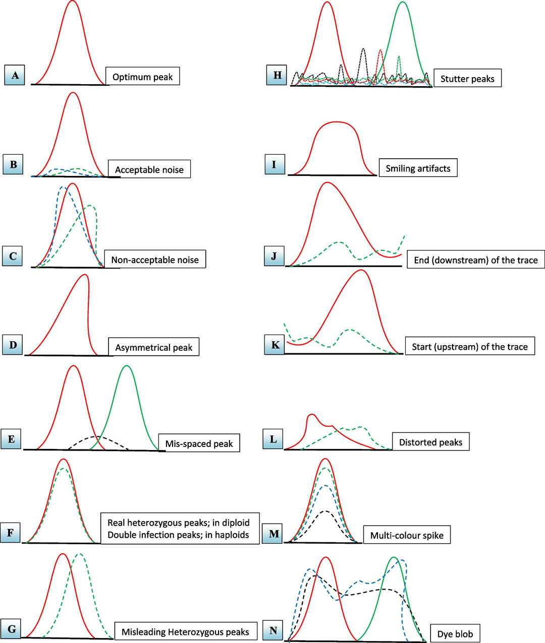 Fig. 1 The main issues encountered in the reading of DNA chromatograms of PCR products based on the Sanger sequencing method.1
The Sanger method was incredibly labor-intensive:
Table 1. Comparison of Sanger's Method with Modern Techniques
| Feature | Sanger Method (DNFB) | Edman Degradation (PITC) | Mass Spectrometry (MS/MS) |
| Principle | N-terminal labeling, full hydrolysis | Sequential N-terminal cleavage | Peptide fragmentation, mass analysis |
| Efficiency/Speed | Very slow, laborious | Moderate, automatable (sequenators) | Very fast, high-throughput |
| Sensitivity | Low (milligram to microgram) | Moderate (nanomole to picomole) | High (picomole to femtomole) |
| Length per Run | 1 amino acid (N-terminal) | Up to ~50 amino acids (automated) | Full peptide sequence (typically 5-30 AA per peptide, many peptides analyzed) |
| Sample Destruction | Complete peptide hydrolysis | N-terminal residue removed, rest intact | Sample consumed in ionization/fragmentation |
| PTM Analysis | Very difficult, indirect | Difficult, some PTMs block Edman | Excellent, can identify and locate PTMs |
| Mixture Analysis | Very difficult, requires pure peptides | Difficult, requires pure peptides | Good, can analyze complex mixtures (LC-MS/MS) |
| Automation | None | Yes (automated sequenators) | Yes (autosamplers, LC, data analysis) |
| Primary Use Today | Historically significant, educational | N-terminal confirmation, specialized | Dominant for most protein sequencing tasks |
Sanger's protein sequencing method not only provided the first glimpse into the precise architecture of proteins but also laid the conceptual and methodological groundwork for decades of subsequent advancements in both protein and nucleic acid sequencing, fundamentally shaping the landscape of modern molecular biology. At Creative Biolabs, we combine decades of experience with cutting-edge technologies and a dedicated team of experts to provide comprehensive de novo sequencing services. We offer de novo antibody sequencing and de novo protein sequencing services, powered by our propriety DASS (Database Assisted Shotgun Sequencing) technology to meet the diverse protein research needs of our clients, driving innovation and advancement in the field of biomedical science.
Learn more about Creative Biolabs' de novo antibody sequencing services:
All listed services and products are For Research Use Only. Do Not use in any diagnostic or therapeutic applications.








