A Comprehensive Overview of Amino Acid Sequencing
Introduction Edman Degradation Mass Spectrometry De Novo Sequencing Applications
Introduction to Amino Acid Sequencing
Definition of Amino Acid Sequencing
The biochemical procedure of amino acid sequencing identifies the exact arrangement of amino acid residues in proteins or peptides. The sequence defines the primary protein structure by providing critical information which guides protein folding and localization to achieve its final function.
Historical Context of Amino Acid Sequencing
The pioneering efforts of Dr. Frederick Sanger established protein sequencing as a scientific field. Dr. Frederick Sanger along with his research team identified the full amino acid sequences of bovine insulin's two polypeptide chains in 1953. Dr. Frederick Sanger received his first Nobel Prize in Chemistry in 1958 for his work which demonstrated that proteins have distinct amino acid sequences and uncovered the primary structure of a protein. The revelation became the basis for contemporary molecular biology and proteomics studies.
Importance in Research and Development
-
Structure-Function Correlation: Protein folding into its functional three-dimensional shape is directed by the sequence of amino acids. Protein sequencing enables scientists to forecast secondary and tertiary structures while identifying functional regions and determining the role of specific residues in protein function.
-
Protein Identification and Characterization: The sequencing process confirms the identity of proteins obtained through isolation or recombinant expression and validates engineered mutations while characterizing new proteins.
-
Elucidating Biological Pathways: The identification of protein sequences involved in specific cellular processes enables scientists to establish interaction networks and comprehend intricate biological systems.
-
Biopharmaceutical Development: Therapeutic proteins require precise sequencing processes for effective characterization as well as maintaining product uniformity and modification detection to meet QC/QA standards.
-
Evolutionary Relationships: Through the comparison of protein sequences across multiple species scientists gain knowledge about evolutionary conservation and divergence which is essential for phylogenetics.
Edman Degradation for Amino Acid Sequencing
Developed by Pehr Edman in the 1950s, Edman degradation remains a valuable technique, particularly for N-terminal sequencing.
Principle of Edman Degradation
This method involves a cyclical chemical process that sequentially removes one amino acid residue at a time from the N-terminus of a peptide. The core steps are:
-
Coupling: Under mildly alkaline conditions, phenyl isothiocyanate (PITC) reacts with the free N-terminal amino group.
-
Cleavage: Under acidic conditions (e.g., anhydrous trifluoroacetic acid - TFA), the N-terminal peptide bond is specifically cleaved, releasing the derivatized N-terminal amino acid as an anilinothiazolinone (ATZ) derivative, leaving the rest of the peptide intact but shortened by one residue.
-
Conversion & Identification: The unstable ATZ-amino acid transforms into the stable phenylthiohydantoin (PTH)-amino acid derivative. High-Performance Liquid Chromatography (HPLC) is used to identify this PTH-amino acid by matching its retention time to established standards.
-
Cycle Repetition: The shortened peptide goes through another round of coupling and cleavage procedures to convert and identify the following amino acid residue.
Strengths and Applications of Edman Degradation
-
Directly determines the N-terminal sequence.
-
Effective for relatively short peptides (typically up to 30-50 residues).
-
Provides unambiguous identification of residues in the initial cycles.
-
Useful for confirming the N-terminus of recombinant proteins or identifying N-terminal modifications that prevent PITC coupling (see Limitations).
Limitations of Edman Degradation
-
Blocked N-termini: If the N-terminal amino group is chemically modified (e.g., acetylated, formylated, or pyroglutamate formation), it cannot react with PITC, preventing sequencing initiation.
-
Decreasing Efficiency: Incomplete reactions and side reactions accumulate over cycles, leading to increased background noise and reduced yield, limiting the practical read length.
-
Sample Requirements: Requires relatively pure samples (µg to mg quantities) and is time-consuming (typically 30-60 minutes per cycle).
-
Inefficient for Mixtures: Not suitable for analyzing complex mixtures of proteins or peptides simultaneously.
-
Destructive: The sample is consumed during the process.
Mass Spectrometry (MS) for Amino Acid Sequencing
The application of mass spectrometry in proteomics transformed protein sequencing through its high sensitivity and throughput capabilities along with complex sample analysis. This method does not sequence proteins from beginning to end in one session like Edman degradation but instead analyzes peptides generated through enzymatic protein digestion, such as with trypsin.
Principle of Mass Spectrometry (MS)
MS determines the mass-to-charge ratio denoted as m/z of ions. Tandem Mass Spectrometry (MS/MS or MS2) stands as the standard method for peptide sequencing.
-
Protein Digestion: The protein of interest undergoes enzymatic cleavage by trypsin to produce smaller peptides through cleavage at the C-terminal end of Arginine (R) and Lysine (K).
-
Separation (Optional but common): The liquid chromatography method Reverse-Phase HPLC (RP-HPLC) usually separates peptide mixtures before they proceed to mass spectrometry analysis.
-
First Mass Analyzer (MS1): The process of ionization allows peptides to separate according to their m/z values and generates a mass spectrum of intact peptide ions that function as precursor ions.
-
Collision-Induced Dissociation (CID): Scientists select the precursor ions they need to study and then fragment them through collisions with inert gas molecules. Fragmentation happens primarily along the peptide backbone. HCD, ETD, and ECD represent alternative fragmentation methods which deliver supplemental data particularly beneficial for PTM analysis.
-
Second Mass Analyzer (MS2): The separation of resulting fragment ions (product ions) occurs through their m/z values to produce a tandem mass spectrum (MS/MS spectrum).
-
Sequence Derivation: The mass differences observed between peaks in the MS/MS spectrum match the masses of individual amino acid residues. Specialized software examines fragment ion patterns which consist of b-ions at the N-terminus and y-ions at the C-terminus to determine the peptide sequence.
Strengths and Applications of Mass Spectrometry (MS)
-
High Sensitivity: The method can perform analysis on quantities ranging from femtomoles (10−15 mol) to attomoles (10 −18 mol).
-
High Throughput: LC-MS/MS systems enable the simultaneous analysis of thousands of peptides during a single experimental run that lasts several hours.
-
Complex Mixtures: This technique enables peptide analysis from complex protein mixtures such as cell lysates.
-
PTM Identification: This technique identifies and localizes post-translational modifications (PTMs) through detection of mass changes in peptides and fragment ions.
-
Internal Sequences: Yields protein sequence data from internal peptide regions beyond the N-terminal sequence.
-
Blocked N-termini: Peptide sequencing remains possible when the protein's N-terminus is blocked if internal peptides are generated.
Limitations of Mass Spectrometry (MS)
-
Indirect Sequencing: Sequences peptides, not the full-length protein directly. Requires bioinformatics tools to assemble peptide sequences into a protein sequence (often aided by a reference database).
-
Sequence Gaps: May not achieve 100% sequence coverage, especially for large proteins or regions resistant to digestion/ionization/fragmentation.
-
Ambiguity: Cannot easily distinguish isobaric residues (Leucine/Isoleucine, having identical mass) without specialized fragmentation techniques (e.g., ECD/ETD) or high-resolution MS. Lysine and Glutamine have very similar masses, requiring high-resolution instruments.
-
Data Complexity: Generates large, complex datasets requiring sophisticated bioinformatics software and expertise for interpretation.
De Novo Sequencing for Amino Acid Sequencing
De novo sequencing involves identifying the sequence of amino acids in a peptide directly from its tandem mass spectrum without using a sequence database.
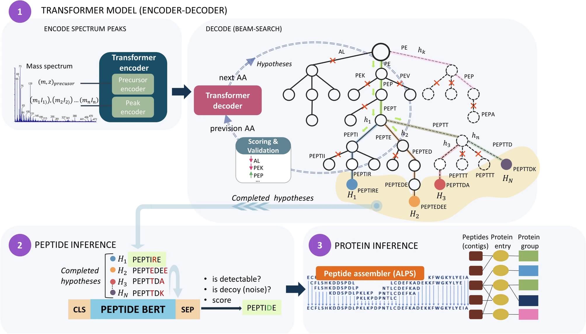 Fig. 1 PowerNovo architecture overview.1
Principle of De Novo Sequencing
The method applies computational algorithms to analyze fragmentation patterns such as b-ions, y-ions, internal ions, and immonium ions in an MS/MS spectrum. The algorithms calculate mass differences between fragment ion peaks to sequentially deduce the amino acid residues. Accurate mass measurements from high-resolution mass spectrometers significantly aid this process.
Strengths and Applications of De Novo Sequencing
-
Database Independent: Essential for sequencing proteins from organisms with unsequenced genomes.
-
Novel Protein Identification: Can identify completely unknown or unexpected proteins/peptides.
-
Antibody Sequencing: It is essential to determine monoclonal antibodies' sequences because variable regions that bind antigens may not have adequate representation in current sequence databases.
-
Sequence Variant Analysis: The Sequence Variant Analysis feature detects genetic variations that reference databases fail to document.
-
Quality Control: Verifying sequences of synthetic peptides or biotherapeutics.
Limitations of De Novo Sequencing
-
Computational Intensity: Requires sophisticated and computationally intensive algorithms.
-
Spectrum Quality Dependent: Heavily reliant on high-quality, information-rich MS/MS spectra.
-
Ambiguity: Prone to errors, especially with low-quality spectra or complex fragmentation patterns. Distinguishing isobaric residues (Leu/Ile) remains challenging.
-
Lower Throughput: De novo interpretation is generally slower and more challenging than database searching.
Table 1. Comparative overview of major amino acid sequencing techniques.
|
Feature
|
Edman Degradation
|
Mass Spectrometry (Database Search)
|
Mass Spectrometry (De Novo)
|
|
Principle
|
Sequential Chemical Degradation
|
MS/MS Fragmentation + Database Match
|
MS/MS Fragmentation + Spectral Interpretation
|
|
Starting Point
|
N-terminus
|
Internal Peptides
|
Internal Peptides
|
|
Read Length
|
Short (<50 residues typical)
|
Peptide length (typically 5-30)
|
Peptide length (typically 5-25)
|
|
Blocked N-Term
|
Fails
|
Tolerated
|
Tolerated
|
|
Sensitivity
|
Moderate (pmol-nmol)
|
High (amol-fmol)
|
High (amol-fmol)
|
|
Throughput
|
Low (sequential)
|
High (LC-MS/MS)
|
Moderate (computationally limited)
|
|
Complex Mixtures
|
Poor
|
Excellent
|
Good (but challenging)
|
|
PTM Analysis
|
Limited / Indirect
|
Excellent
|
Good (requires careful interpretation)
|
|
Primary Use
|
N-terminal confirmation, Short peptides
|
Protein ID, PTMs, Quantification
|
Novel proteins, Antibodies, Sequence Variants
|
|
Database Req.
|
No
|
Yes
|
No
|
|
Key Limitation
|
Length, Blocked N-Term, Speed
|
Sequence Gaps, Database Dependency
|
Accuracy, Isobaric Residues, Complexity
|
Applications of Amino Acid Sequencing in Research
Protein Identification and Characterization
The fundamental process in protein research involves confirming the identity of proteins. Researchers utilize MS-based sequencing along with Peptide Mass Fingerprinting to determine which proteins have been isolated from biological samples by comparing experimental peptide masses and sequences to theoretical database values. Researchers must verify the sequence of recombinantly expressed proteins for research applications and biotherapeutic development.
Analysis of Post-Translational Modifications (PTMs)
Protein synthesis produces amino acid side chains that undergo covalent modifications known as PTMs which significantly increase the proteome's functional capabilities. Protein activity and behavior including location and interaction patterns as well as degradation rates are regulated through post-translational changes such as phosphorylation, glycosylation, ubiquitination, methylation, acetylation, and disulfide bond formation. The primary technique for determining PTM types and their positions on polypeptide chains is MS-based sequencing which utilizes characteristic mass shifts on peptides and their fragments for detection.
Discovery of Disease Biomarkers
An analysis of protein profiles from healthy and diseased tissues or biofluids identifies proteins that undergo significant changes in abundance or modification status. Quantitative mass spectrometry techniques enable scientists to sequence and identify proteins that show differential expression or peptide modifications. These proteins hold promise as research biomarkers for detecting disease and monitoring therapeutic responses but need additional validation before they can be used in clinical applications. Studies of cancer research models through sequencing techniques reveal altered PTM patterns and specific protein isoforms that help to understand tumorigenesis.
Drug Development and Biologics Characterization
The pharmaceutical industry relies on amino acid sequencing for essential operations.
-
Target Validation: The process of identifying the correct amino acid sequence of a potential drug target protein.
-
Biotherapeutic Development: Therapeutic proteins including monoclonal antibodies (mAbs), insulin, growth factors and vaccines require accurate and complete sequence data for characterization and regulatory submissions. Characterization requires validation of mAbs variable regions alongside detection of PTMs such as glycosylation patterns which influence mAbs efficacy and immunogenicity as well as identification of sequence variants or degradation products.
-
Quality Control (QC): Biopharmaceutical products maintain batch-to-batch consistency through primary structure confirmation and detection of modifications and impurities.
-
Epitope Mapping: Scientists determine the exact amino acid sequence, known as the epitope, which an antibody identifies on an antigen.
Amino acid sequencing has evolved dramatically from the pioneering work of Sanger to the sophisticated mass spectrometry-driven approaches widely used today. At Creative Biolabs, we leverage state-of-the-art sequencing technologies and extensive expertise to provide robust and accurate amino acid sequencing services. We offer de novo antibody sequencing and de novo protein sequencing services, powered by our propriety DASS (Database Assisted Shotgun Sequencing) technology to meet the diverse protein research needs of our clients, driving innovation and advancement in the field of biomedical science.
Learn more about Creative Biolabs' de novo antibody sequencing services:
Reference
-
Petrovskiy, Denis V., et al. "PowerNovo: de novo peptide sequencing via tandem mass spectrometry using an ensemble of transformer and BERT models." Scientific Reports 14.1 (2024): 15000. Distributed under Open Access license CC BY 4.0, without modification. https://doi.org/10.1038/s41598-024-65861-0
All listed services and products are For Research Use Only. Do Not use in any diagnostic or therapeutic applications.









