
Creative Biolabs is offering the most comprehensive services for antibody development projects. With strict regulation and effective execution, we are dedicated to providing the most valuable solutions to complete your projects.
HighlightsCreative Biolabs is offering the most comprehensive services for antibody development projects. With strict regulation and effective execution, we are dedicated to providing the most valuable solutions to complete your projects.
HighlightsCreative Biolabs has established a broad range of platforms for developing novel antibodies or equivalents. These cutting-edge technologies enable our scientists to meet your demands from different aspects and tailor the most appropriate solution that contributes to the success of your projects.
HighlightsWith deep understanding in antibody-related realms and extensive project experience, Creative Biolabs offers a variety of references to help you learn more about our capacities and achievements, including infographic, flyer, case study, peer-reviewed publications, and all kinds of knowledge that can assist your projects. You are also welcome to contact us directly for more specific solutions.
HighlightsGet a real taste of Creative Biolabs, one of the most professional custom service providers in the world. We are committed to providing highly customized comprehensive solutions with the best quality to advance your projects.
HighlightsDe novo peptide sequencing is a sophisticated analytical approach used to determine the primary amino acid sequence of a peptide directly from its tandem mass spectrometry (MS/MS) fragmentation data, without relying on existing protein databases or genomic sequences.
This technique is indispensable in various research domains for several critical reasons:
The journey of peptide sequencing has evolved significantly over the past century. Early methods, such as Edman degradation, developed by Pehr Edman in the 1950s, were revolutionary. This sequential chemical cleavage method allowed for the removal and identification of one amino acid at a time from the N-terminus of a peptide. While groundbreaking, Edman degradation was labor-intensive, required relatively large sample amounts, and was limited to peptides of approximately 50-60 amino acids due to accumulating errors.
The advent of mass spectrometry (MS) in the latter half of the 20th century marked a paradigm shift. The development of soft ionization techniques like Electrospray Ionization (ESI) and Matrix-Assisted Laser Desorption/Ionization (MALDI) in the late 1980s enabled the analysis of large, non-volatile biomolecules like peptides and proteins without significant fragmentation. This paved the way for tandem mass spectrometry (MS/MS), which became the cornerstone of modern de novo sequencing, offering higher sensitivity, speed, and the ability to analyze complex mixtures.
The core of de novo peptide sequencing lies in the intelligent interpretation of MS/MS fragmentation data.
Tandem mass spectrometry involves multiple stages of mass analysis, typically separated by a fragmentation step. The general workflow for peptide sequencing using MS/MS is as follows:
The method used to fragment peptides significantly influences the types of ions generated and the resulting MS/MS spectrum. Common fragmentation techniques include:
When a peptide fragments, it typically breaks along its backbone, generating characteristic ion series. These ions are named based on the site of cleavage and whether they retain the N-terminal or C-terminal part of the original peptide.
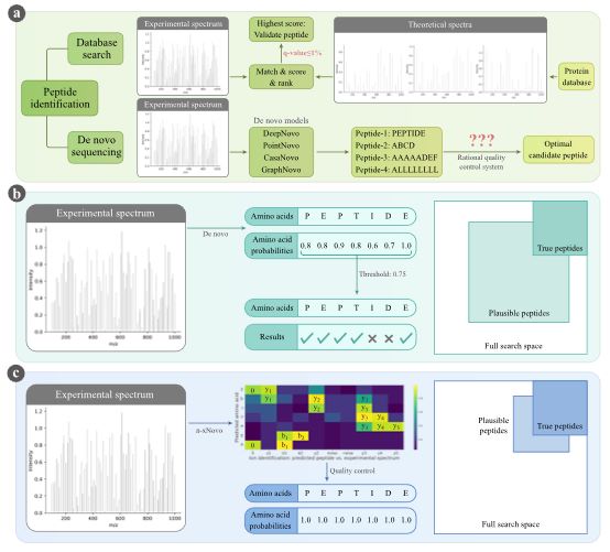 Fig. 1 Schematic overview of peptide identification.1
The process of de novo sequencing involves computationally reconstructing the peptide sequence from the observed fragment ions. The fundamental principle is that the mass difference between consecutive fragment ions corresponds to the mass of a single amino acid residue. By identifying these mass differences and matching them to the known masses of all 20 standard amino acids, the sequence can be deduced.
One of the primary advantages of de novo sequencing is its ability to characterize peptides and proteins for which no sequence information exists in public databases. This is particularly relevant for:
PTMs are crucial for regulating protein function, localization, and interaction. However, many PTMs are dynamic and context-dependent, making their identification challenging through database searching alone. De novo sequencing excels here because it does not require prior knowledge of the PTM. By observing unexpected mass shifts between adjacent fragment ions, researchers can infer the presence and exact location of modifications.
For organisms whose genomes have not yet been sequenced, traditional proteomics approaches relying on database matching are ineffective. De novo peptide sequencing provides the only means to directly characterize their proteomes, offering insights into their biology, metabolism, and unique adaptations. This is particularly valuable in environmental proteomics, microbial ecology, and biodiversity studies.
While database search engines are highly efficient for identifying peptides from known protein databases, they are limited by the completeness and accuracy of those databases. De novo sequencing serves as a powerful complement:
The accuracy of de novo sequencing is highly dependent on the quality of the MS/MS spectrum.
As mentioned previously, the existence of isobaric amino acids (e.g., Leucine/Isoleucine, Lysine/Glutamine) poses a significant challenge. Without additional information or high-resolution data that can differentiate their subtle mass differences (e.g., using ultra-high resolution FT-ICR MS), de novo algorithms often cannot distinguish between these residues, leading to sequence ambiguities. This often results in reporting "L/I" or "K/Q" in the sequence.
For a given set of fragment ions, it can sometimes be difficult to definitively determine the N-terminal to C-terminal direction of the peptide. While the presence of both b- and y-ion series typically helps establish directionality, in cases of sparse fragmentation or when only one ion series is dominant, the orientation of the sequence can be ambiguous. Advanced algorithms often employ strategies like analyzing the precursor ion's charge state and the presence of specific diagnostic ions to infer directionality.
As peptide length increases, the number of possible fragment ions grows exponentially, and the complexity of the MS/MS spectrum increases. This significantly escalates the computational resources and time required for de novo algorithms to accurately reconstruct the sequence. Longer peptides also tend to fragment less completely, exacerbating the problem. For very long peptides, it is often necessary to digest them into smaller, more manageable fragments before sequencing.
While de novo sequencing has made significant strides, it still faces limitations in achieving 100% accuracy and full sequence coverage, especially for complex or modified peptides.
De novo peptide sequencing stands as a powerful and indispensable tool in the modern proteomics landscape. At Creative Biolabs, we offer de novo antibody sequencing and de novo protein sequencing services, powered by our propriety DASS (Database Assisted Shotgun Sequencing) technology to meet the diverse protein research needs of our clients, driving innovation and advancement in the field of biomedical science.
Learn more about Creative Biolabs' de novo antibody sequencing services:
All listed services and products are For Research Use Only. Do Not use in any diagnostic or therapeutic applications.








