
Creative Biolabs is offering the most comprehensive services for antibody development projects. With strict regulation and effective execution, we are dedicated to providing the most valuable solutions to complete your projects.
HighlightsCreative Biolabs is offering the most comprehensive services for antibody development projects. With strict regulation and effective execution, we are dedicated to providing the most valuable solutions to complete your projects.
HighlightsCreative Biolabs has established a broad range of platforms for developing novel antibodies or equivalents. These cutting-edge technologies enable our scientists to meet your demands from different aspects and tailor the most appropriate solution that contributes to the success of your projects.
HighlightsWith deep understanding in antibody-related realms and extensive project experience, Creative Biolabs offers a variety of references to help you learn more about our capacities and achievements, including infographic, flyer, case study, peer-reviewed publications, and all kinds of knowledge that can assist your projects. You are also welcome to contact us directly for more specific solutions.
HighlightsGet a real taste of Creative Biolabs, one of the most professional custom service providers in the world. We are committed to providing highly customized comprehensive solutions with the best quality to advance your projects.
HighlightsCreative Biolabs provides specialized IHC-positive antibody discovery services, focused on generating high-quality antibodies optimized for IHC applications. We leverage advanced technologies and proven methodologies to deliver tailored solutions, empowering our clients to achieve accurate and reliable results.
Immunohistochemistry (IHC) is a fundamental technique for visualizing and precisely localizing specific proteins within tissue sections. This method is predicated on the highly specific binding interaction between an antibody and its cognate antigen. In essence, IHC involves applying antibodies to tissue samples, where they bind to target proteins. Subsequent detection methods, often involving enzymatic reactions or fluorescent labels, allow for the visualization of this binding, thus revealing the distribution and expression patterns of the protein of interest.
The mechanism of action in IHC centers on the antibody's capacity to specifically recognize and bind to a specific region on the target protein, known as the epitope. Primary antibodies, which directly target the protein, are initially applied to the tissue. To enhance signal detection, secondary antibodies, which bind to the primary antibodies, are often employed. When the appropriate substrate is added, enzymatic reporters produce a colored precipitate, while fluorophores emit light, allowing for microscopic visualization of the target protein's location within the tissue.
IHC holds a crucial role in various fields, including disease diagnosis, prognostic and predictive marker identification, research investigations, and drug discovery. The technique's capacity to provide spatial and morphological context to protein expression makes it indispensable for understanding complex biological processes and disease mechanisms.
IHC hinges upon the precise detection of antibodies directed against specific epitopes, thereby imposing heightened requisites on antibody generation. Within our hybridoma screening framework, Creative Biolabs meticulously cultivates IHC-specific antibodies through dedicated IHC-positive hybridoma screening. Leveraging a sophisticated and exhaustive hybridoma platform, we extend a spectrum of comprehensive hybridoma experimental services tailored to your needs.
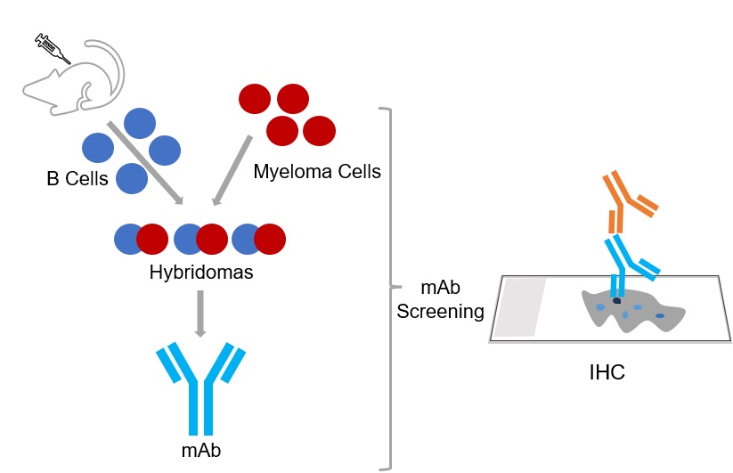
The foundation of successful antibody development lies in the careful design and preparation of the immunogen. Our team collaborates closely with clients to determine the most suitable antigen, which may encompass recombinant proteins, synthetic peptides, or fixed tissue samples. We employ advanced techniques to ensure the antigen is presented in an optimal conformation to elicit the desired antibody response.
This process involves a series of carefully timed immunogen administrations to stimulate the animal's immune system to produce antibodies against the target antigen. We closely monitor the animal's response and adjust the protocol as needed to maximize antibody production.
Once the animal has developed a robust immune response, antibody-producing B cells are isolated and subjected to fusion with myeloma cells to create hybridomas. These hybridomas are immortalized cells that have the capability to continuously produce monoclonal antibodies. This crucial step allows for the generation of a sustainable source of antibodies with the desired specificity.
A critical aspect of our service is the implementation of IHC-specific screening assays. We develop and optimize assays that directly evaluate the performance of the generated antibodies in tissue sections. This rigorous screening process ensures that only antibodies demonstrating optimal staining characteristics, including high specificity and sensitivity, are selected for further development.
Hybridomas that exhibit promising IHC staining patterns undergo further characterization to assess their antibody production levels, specificity, and stability. We employ a range of techniques, such as ELISA, Western blotting, and flow cytometry, to comprehensively evaluate the antibodies and select the clones that best meet the project's requirements.
Selected hybridoma clones are then cultured to produce larger quantities of the desired antibodies. We utilize state-of-the-art cell culture systems to ensure efficient and scalable antibody production. The antibodies are subsequently purified using advanced techniques to remove any contaminants and obtain highly purified antibody preparations.
We perform rigorous validation and quality control testing on the final antibody products. This process includes testing in various tissue types, comparing with known positive and negative controls, and assessing staining patterns. Our commitment to quality ensures that our clients receive antibodies that are reliable, reproducible, and suitable for their specific IHC applications.
In the realm of IHC, elucidating the specificity of antibodies poses a formidable challenge, primarily due to the scarcity of ideal controls. Wherein, gene-knockout tissues or specific transgenic fusion proteins serve as viable references for staining. Consequently, an ancillary assay, such as ELISA, Western Blotting (WB), or IP, becomes imperative to corroborate the antibody's binding specificity to the target antigen. Subsequently, the antibody undergoes scrutiny in IHC assays, demanding specificity in staining—manifested by positivity in select tissues or cells, with staining intensity surpassing that of isotype-matched negative control antibodies. To obtain antibodies suitable for IHC, it is essential to conduct an initial screening using ELISA, Western blotting (for epitopes in denatured conditions), and/or immunoprecipitation (for epitopes in native conditions). Empirical evidence suggests a high likelihood of obtaining IHC-positive clones when multiple ELISA/WB/IP-positive clones are secured.
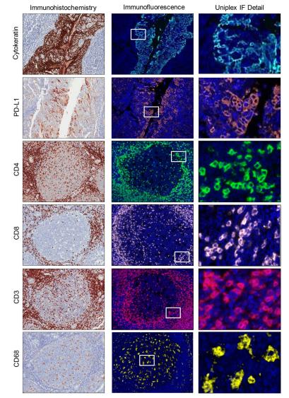 Fig.1 Microphotographs of representative examples of validation from IHC and uniplex IF.1
Parra, Edwin R., et al. conducted IHC analysis utilizing a chromogen-based approach with an automated staining system1. By analyzing the staining intensity, ranging from negative to positive in control samples, and assessing the uniformity of staining across various antibodies targeting specific cell expressions, they determined the appropriate antibody titer for achieving the correct staining pattern in IHC. Furthermore, they evaluated various markers using both chromogenic IHC and uniplex immunofluorescence (IF) methods to ensure consistency in staining patterns between the two immunohistochemical techniques.
Creative Biolabs delivers antibodies of exceptional specificity:
A: IHC is a method that employs antibodies to identify particular proteins in tissue samples. It's a vital tool in medical research and clinical diagnostics because it allows scientists and doctors to see where proteins are located within cells and tissues. This information is vital for understanding how diseases develop, diagnosing conditions like cancer, and developing new treatments.
A: Developing antibodies for IHC is more complex than for other techniques due to several factors. Tissue processing can alter the shape of proteins, making it difficult for antibodies to recognize them. IHC also requires antibodies to be highly specific to avoid binding to other proteins in the tissue, which would lead to inaccurate results. Additionally, the antibodies need to penetrate the tissue effectively and remain stable throughout the IHC process.
A: We employ several strategies to ensure high specificity. We start with careful antigen design and preparation. Then, we use stringent screening processes, including IHC-specific assays, to select antibodies that bind only to the target protein in tissue sections. We also perform extensive validation testing, including comparing staining in positive and negative control tissues, to confirm specificity.
A: We can use a variety of antigens, depending on the specific project requirements. Common antigen types include recombinant proteins, synthetic peptides, and even denatured antigens, such as fixed tissue samples. The choice of antigen is crucial and is tailored to elicit the most effective antibody response for IHC applications. We consider factors such as protein structure and potential modifications during tissue processing.
A: IHC-positive hybridoma screening is a specialized screening process we use to identify hybridoma clones that produce antibodies that work well in IHC. Unlike general antibody screening, this method directly tests the antibodies' performance on tissue sections. We also use tissue microarrays for hybridoma screening. This ensures that we select only antibodies that can effectively bind to the target protein in its native tissue context.
A: We can work with a wide range of tissue samples, including formalin-fixed, frozen tissue, and paraffin-embedded (FFPE) tissue sections. The specific type of tissue sample used for testing will depend on the client's research or diagnostic needs. We also optimize our protocols to ensure that the antibodies perform well with the client's specific tissue types.
Endowed with extensive proficiency in mAb production, Creative Biolabs emerges as a venerable leader in this field. Supported by our state-of-the-art hybridoma facilities, we offer a comprehensive suite of hybridoma-based services. Should our offerings pique your interest, we wholeheartedly invite you to contact us for additional clarification.
All listed services and products are For Research Use Only. Do Not use in any diagnostic or therapeutic applications.








