
Creative Biolabs is offering the most comprehensive services for antibody development projects. With strict regulation and effective execution, we are dedicated to providing the most valuable solutions to complete your projects.
HighlightsCreative Biolabs is offering the most comprehensive services for antibody development projects. With strict regulation and effective execution, we are dedicated to providing the most valuable solutions to complete your projects.
HighlightsCreative Biolabs has established a broad range of platforms for developing novel antibodies or equivalents. These cutting-edge technologies enable our scientists to meet your demands from different aspects and tailor the most appropriate solution that contributes to the success of your projects.
HighlightsWith deep understanding in antibody-related realms and extensive project experience, Creative Biolabs offers a variety of references to help you learn more about our capacities and achievements, including infographic, flyer, case study, peer-reviewed publications, and all kinds of knowledge that can assist your projects. You are also welcome to contact us directly for more specific solutions.
HighlightsGet a real taste of Creative Biolabs, one of the most professional custom service providers in the world. We are committed to providing highly customized comprehensive solutions with the best quality to advance your projects.
HighlightsCreative Biolabs provides a Mitophagy Analysis Service to address key challenges in metabolic and cellular research, including difficulty quantifying mitochondrial turnover, distinguishing selective mitophagy from general autophagy, and identifying the threshold at which metabolic stress leads to irreversible cellular damage. This service enables high-resolution, quantitative analysis of mitochondrial degradation and integrity through advanced fluorescence co-localization, mitophagy flux assays, and targeted mapping. By delivering clear, mechanism-driven insights, Creative Biolabs supports efficient evaluation of candidate compounds and accelerates the understanding of cellular responses under metabolic stress.
Mitophagy is a selective autophagic process responsible for removing damaged or dysfunctional mitochondria, playing a vital role in maintaining cellular homeostasis. Studies have demonstrated that pathways such as PINK1/Parkin-mediated signaling regulate mitochondrial turnover and are closely linked to diseases, including neurodegeneration and cancer. Advances in fluorescence imaging, biosensor development, and molecular biology techniques have significantly improved the ability to monitor mitophagy dynamics, supporting its growing importance as both a research focus and therapeutic target.
Creative Biolabs provides an analytical suite designed to decode the complexities of mitochondrial quality control. We don't just provide raw data; we deliver biological context. Our service allows you to determine exactly how a therapeutic agent influences the clearance of damaged mitochondria, which is critical for projects targeting neurodegeneration, cardiovascular disease, and retinal integrity. By integrating our findings, you can confidently validate drug targets and optimize dosing based on cellular self-cleaning efficiency. We provide:
Our integrated approach ensures reliable data generation, reduced experimental variability, and actionable biological insights. Discover How We Can Help – Request a Consultation.
| Fluorescence Imaging-Based Analysis | Biosensor and Reporter Systems |
|
|
| Protein and Molecular Assays | Functional Mitochondrial Readouts |
|
|
Required Starting Materials:
Clients typically provide well-characterized biological samples (e.g., cultured cell lines, such as cancer or primary cells), treatment conditions (e.g., small molecules or genetic modulators), and experimental background information (e.g., target pathways or expected outcomes).
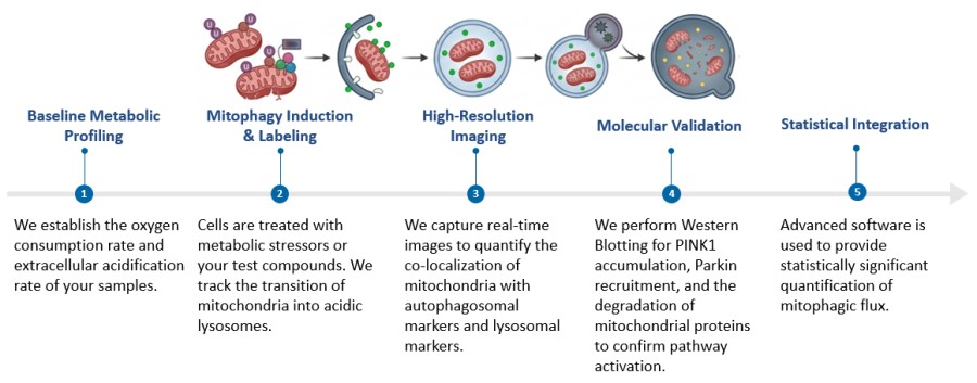
You will receive a comprehensive technical report including:
(1) High-resolution, publication-ready confocal micrographs with scale bars and annotations
(2) Quantitative flux graphs comparing control vs. treated groups
(3) A detailed summary of the PINK1-Parkin axis response.
Creative Biolabs offers a world-class Mitophagy Analysis Service that delivers the precision and biological insight required for modern drug discovery. Our comprehensive solutions cover mitophagic flux quantification, mitochondrial and lysosome co-localization analysis, and detailed mapping of the PINK1-Parkin signaling axis. We integrate advanced fluorescence imaging, biosensor-based assays, protein expression profiling, and functional mitochondrial readouts such as membrane potential, ROS, and ATP levels. With flexible experimental design and multi-platform validation, our service helps you overcome technical bottlenecks and generate reliable, publication-ready data to accelerate your path to the clinic. Contact Our Team for More Information and to Discuss Your Project.
All listed services and products are For Research Use Only. Do Not use in any diagnostic or therapeutic applications.








