Overview Service Features Published Data FAQs Scientific Resources Related Services
Creative Biolabs provides high-efficiency iPSC generation and application services for our customers all over the world. With the great potential in drug discovery, scientists at Creative Biolabs have developed the multi-electrode array (MEA) technology for the electrophysiological characterization of iPSC-derived cell lines, including cardiomyocytes and neurons.
Overview of Electrophysiological Characterization for iPSC
Introduction of Multi-electrode Array (MEA)
Multi-electrode arrays (MEAs) are devices comprised of multiple plates or shanks through which neural signals are obtained and delivered. In general, there are two types of MEAs which are implantable MEAs used in vivo and non-implantable MEAs used in vitro. In biological research, MEA has been used for the electrophysiological characterization of iPS cell lines. Compared with the traditional patch-clamp methods, MEA provides a rapid and efficient approach for the analysis of electrophysiological characterization with higher throughput and reduced operator skill requirement. Moreover, MEA also enables experimental protocols including long-term chronic drug exposure, both 2D and 3D cultures for mixed cell populations, and the analysis of drug impact on cell-to-cell transmission of electrical signaling that is precluded by standard patch-clamp methodology. In this case, the combination of MEA and iPSC-derived cells provide an attractive approach for drug discovery.
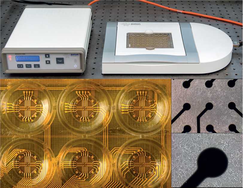
 Fig.1 Experimental setup for measurements of spontaneous electrical network activity.1
Electrophysiological Characterization of iPSC-derived Cardiomyocytes
In recent years, the production of cardiomyocytes (CM) derived from iPSCs shows promising potential for drug safety assessment. Now we are able to use MEA analysis of iPSC-CM to generate multiparameter data to profile drug impact on cardiomyocyte electrophysiology using a panel of various compounds active against key cardiac ion channels. Thus, the drug liability can be evaluated early in the drug discovery process. The main protocol and content list as follow:
-
Molecular and cellular characterization of hiPSC-CMs.
-
Validation of MEA culture.
-
Identification of pacemaking cells in the monolayer cardiac sheet.
-
Characterization of cardiac maturity and conduction velocity.
Electrophysiological Characterization of iPSC-derived Neurons
Current traditional test for neurotoxicity always relies on expensive and time-consuming in vivo animal experiments which limit its wide applications. The rapid development of iPSC technology presents great potential to solve this problem. The immunofluorescent staining would be used to demonstrate the human iPSC-derived neurons from various origins. According to the use of multi-well microelectrode array (mwMEA) recordings, we can demonstrate these human iPSC-derived cultures developing spontaneous neuronal activity over time and it can be modulated by different physiological, toxicological and pharmacological compounds. Upon further electrophysiological characterization and toxicological validation, iPSC-based models can facilitate predictions of neurotoxicity.
In addition, Creative Biolabs provides other various electrophysiological characterization services for different iPSC-derived cell types based on our advanced MEA technology. With years of experience, we are capable of meeting the specific need of each customer with the best project design. If you are interested in our services, please do not hesitate to contact us for more details.
Services at Creative Biolabs
MEA is a non-invasive, label-free approach that offers the advantage of real-time, high-throughput screening. Our company uses advanced MEA technology to record and analyze the electrical activity of iPSC-derived neurons and cardiomyocytes.
Our services comprise the following key features:
-
Source of iPSCs: We work with client iPSC lines or alternatively generate iPSCs from somatic cells provided by clients, utilizing cutting-edge protocols and technologies. These cells can be differentiated into specific cell types, such as neurons or cardiac muscle cells, based on the research needs.
-
iPSC Culture and Differentiation: We master the cultivation of iPSCs and the differentiation of these cells into cardiomyocytes, neurons, or other cell types. Our team is experienced in maintaining the pluripotency of iPSCs and directing their differentiation into defined cell types using validated protocols.
-
Electrophysiological Characterization: Using MEA, we characterize the electrophysiological properties of iPSC-derived cells. For iPSC-derived cardiomyocytes, we measure parameters such as beat rate, field potential duration, QT intervals, etc., that reflect cardiac function. For iPSC-derived neurons, we can record and analyze spikes and bursts that indicate neuronal network activity.
-
Data Analysis and Reporting: Our team provides comprehensive data analysis and reporting. Each report will include information on the study design, methodology, raw and analyzed data, statistical analysis and conclusion. Our detailed report will provide you with a complete understanding of the test results.
It should be noted that all of our iPSC and MEA services are strictly for research use only and not for use in diagnostic or therapeutic procedures. At all stages, we work under strict ethical guidelines and follow necessary protocols to responsibly handle and process stem cells.
Features of Our Services
-
Comprehensive Analysis - Our services provide a comprehensive analysis of functional properties of various iPSC-derived cell types, including neurons, cardiomyocytes, and other excitable cells.
-
Cutting-Edge Technologies - These allow for accurate and precise measurement of key electrophysiological properties including membrane potential, action potential amplitude and duration, spontaneous firing rate, and synaptic activity.
-
High-Throughput Screening - This means we're able to assess the effects of hundreds to thousands of different variables or compounds on the electrophysiological properties of iPSC-derived cells in a time-efficient manner.
-
Service Flexibility - We offer flexible service models, from fee-for-service to full collaboration, ensuring that our clients can choose the model that best fits their budget and research needs.
Overall, our electrophysiological characterization services not only facilitate superior research standards but also significantly cut-down on research time, manpower, and resources, thereby accelerating research progress.
Published Data
Below are the findings presented in the article related to electrophysiological characterization of iPSC.
Researchers have used HD-MEA to characterize and compare human induced pluripotent stem cell-derived dopaminergic and motor neurons in vitro. Reproducible electrophysiological networks, single-cell and subcellular metrics for phenotypic characterization and drug testing.
In order to compare the electrophysiological properties of the four human iPSC-derived neuronal cell lines, they first examined and compared mean firing rate (MFR), mean spike amplitude (MSA), mean ISI coefficient of variation (ISIcv), and percentage of active electrodes (pAE). It was found that the average firing rates of both motor (hMN) and dopaminergic (hDN) neuronal lines increased significantly from DIV 7 to DIV 21. The hMN had a higher MFR, MSA, and pAE compared to the hDN.
FAQs
-
Q: What type of equipment do you use in these services?
A: We employ state-of-the-art electrophysiological equipment and tools for precision and accuracy in our services. This includes patch clamp systems for studying ion channels, microelectrode arrays for recording activity from multiple cells simultaneously, and imaging systems for assessing cellular morphology and changes.
-
Q: Can you provide assistance with optimizing the differentiation protocols to generate iPSC-derived neurons with specific electrophysiological properties?
A: Yes, we offer consultancy services to optimize differentiation protocols for generating iPSC-derived neurons with desired electrophysiological properties. Our team can provide guidance on culture conditions, growth factors, and small molecules to induce the differentiation of iPSCs into functional neurons.
-
Q: How do you handle unexpected variability or deviations in electrophysiological recordings during the course of the experiment?
A: We have protocols in place to address unexpected variability or deviations in electrophysiological recordings promptly. Our team employs rigorous quality control measures and data analysis techniques to identify and mitigate any anomalies, ensuring the integrity and reliability of the experimental data. Additionally, we collaborate closely with customers to troubleshoot issues and implement corrective measures as needed.
-
Q: What are the costs associated with utilizing your electrophysiological characterization services for iPSC experiments?
A: Our pricing structure is tailored to accommodate different budgetary constraints and project scopes. Costs may vary depending on factors such as the complexity of the experiment, the number of samples, and additional services required. We provide transparent pricing estimates and work closely with customers to develop a cost-effective solution that meets their needs.
Scientific Resources
Reference
-
Tukker, Anke M., et al. "Is the time right for in vitro neurotoxicity testing using human iPSC-derived neurons?." ALTEX-Alternatives to animal experimentation 33.3 (2016): 261-271. Distributed under Open Access license CC BY 4.0, without modification.
For
Research Use Only. Not For Clinical Use.
 Fig.1 Experimental setup for measurements of spontaneous electrical network activity.1