Overview Service Features Published Data FAQs Scientific Resources Related Services
Based on our extensive experience and advanced platform, Creative Biolabs has been a long-term expert in the field of iPSC technology. Now we provide the morphological analysis service for iPSCs during induced differentiation and reverse programming.
Overview of Morphology of iPSC
Introduction of iPSC Morphology
Obtained through the iPSC reprogramming of adult or fetal differentiated somatic cells by ectopic expression of a set of core pluripotency-related transcription factors, iPSCs present great therapeutic potential both in biological research and drug discovery. However, a fundamental problem concerning different degrees of induced pluripotency during the reprogramming process arises. At present, the pluripotency characteristics of human IPSCs are evaluated by pluripotency-associated cell surface markers according to molecular criteria and functional aspect by teratoma assays. In order to evaluate the morphological evolution of cells, an analysis is essential to obtain the fine structure of human iPSCs compared with human embryonic stem cells (hESCs) and differentiated cells. Now we are able to analyze the morphology of iPSC by confocal microscopy.
Analysis of iPSC Morphology
During our assay, there are three cell types have been used: human iPSCs, human ESCs, and mesenchymal stem cells (MSCs). Human ESCs can differentiate into MSCs. In this case, we are able to determine the ultrastructural characteristics of human iPSCs compared with their differentiated state in the same culture condition based on our reversible differentiation methodology. When cultured in presence of MEFs, we have found that the particular characteristics and morphological organization of the iPSC colonies are similar to those of inner mass cells at the human blastocyst stage of development, mouse embryoid bodies, and human ESCs. Moreover, we have found that the colonies contact with the culture condition via the microvillar side of the epithelial cells and there are large intercellular spaces and internal ducts to allow the transport of medium.
Services at Creative Biolabs
Our service focuses on providing detailed characterization and analysis of induced pluripotent stem cells. Here's a comprehensive description of what this service typically includes:
-
Cell Culture and Maintenance
-
We culture iPSCs using validated protocols to maintain pluripotency and quality.
-
Routine passaging and monitoring to ensure healthy cell growth.
-
Morphological Assessment
-
Regular examination of iPSC morphology under phase contrast microscopy to assess cell density, colony morphology, and overall health.
-
Monitoring iPSCs in real-time to observe growth patterns, colony formation, and any signs of differentiation.
-
Using staining methods to evaluate cell viability and proliferation rates.
-
Quality Control Measures
-
Immunocytochemistry (ICC) or flow cytometry analysis to confirm expression of pluripotency markers such as Oct4, Sox2, Nanog, SSEA-4, and Tra-1-60/81.
-
Assessing iPSCs for chromosomal stability and to detect any abnormalities.
-
Ensuring cultures are free from microbial contamination.
-
Differentiation Potential Testing
-
Evaluating iPSCs' ability to differentiate into cells of all three germ layers (ectoderm, mesoderm, and endoderm) under appropriate conditions.
-
In vivo testing in immunocompromised mice to confirm iPSCs' pluripotency and capacity to form tissues from all germ layers.
-
Additional Services
-
Cas protein-mediated genome editing for creating iPSC lines with specific genetic modifications.
Our iPSC morphology service is intended for research purposes only, providing essential characterization data for researchers studying disease mechanisms, drug discovery, and regenerative medicine.
Main Protocol for Our Analysis
-
Generation and culture of iPSCs, ESCs, and MSCs.
-
Preparation of supernatant particles from cultures.
-
Transmission electron microscopy.
-
Structure analysis of iPSCs, ESCs, and MSCs.
With our well-established iPSC platform, Creative Biolabs is dedicated to helping you with your special project. We are confident in offering iPSCs morphology analysis service for our customers all over the world. On the basis of provided cells, we will start with the cell culture and finally obtain the results through electron microscopy. Creative Biolabs also provides other various services regarding iPSC generation and applications. Please feel free to contact us for more details.
Features of Our Services
-
Comprehensive Characterization: Gain detailed insights into the morphology of your iPSC colonies, crucial for assessing pluripotency and culture quality.
-
Data-Driven Decisions: Use quantitative data to inform decisions regarding experimental protocols or cell line optimization.
-
Customization: Tailor the service to meet specific research requirements or regulatory standards.
-
Expert Support: Access to our team of experienced scientists who can provide guidance and support throughout the process.
Published Data
Below are the findings presented in the article related to morphology of iPSC.
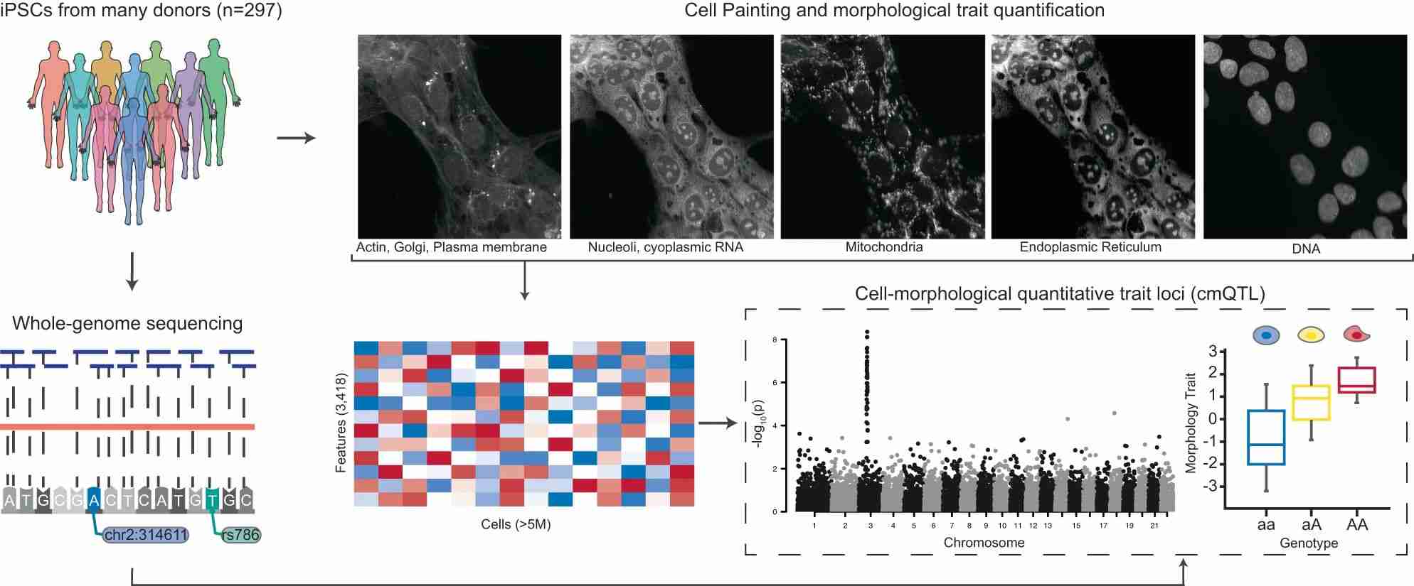
Similar to molecular phenotyping, morphological analysis provides insight into the function of genes and variants. Matthew Tegtmeyer, et al. combined genome sequencing and high-content imaging methods on iPSCs from 297 unique donors to investigate the relationship between genetic variation and cell morphology.
They expanded iPSC lines from 297 donors, performed quality control checks, and then performed cell-coating high-throughput imaging and 30X whole genome sequencing (WGS). They quantified 3418 morphological features per cell using associated software. They inferred genetic variants from the WGS data and investigated whether individual morphological features were associated with rare and common variants.
 Fig. 1 Morphological profiling and whole-genome sequencing on iPSCs.1
FAQs
-
Q: What types of morphology studies can be conducted using iPSCs?
A: iPSCs are versatile tools for studying various aspects of cellular morphology. Researchers can investigate cell shape, size, organelle structure, and distribution using advanced microscopy techniques. Moreover, iPSCs differentiated into specific lineages allow for studying morphological changes during development or disease progression. This flexibility makes iPSCs invaluable for exploring fundamental biological processes and modeling human diseases at the cellular level.
-
Q: What equipment and facilities are available for morphology studies using iPSCs?
A: Our facility is equipped with state-of-the-art microscopy systems capable of high-resolution imaging and live-cell analysis. We provide controlled environments for iPSC culture, maintaining optimal conditions for cell growth and differentiation. Advanced imaging software allows for detailed morphometric analyses and 3D reconstructions of cellular structures. These resources support comprehensive morphology studies, ensuring accurate and insightful data acquisition for researchers engaged in iPSC-based investigations.
-
Q: Can you provide examples of previous morphology studies using iPSCs from your service?
A: Our research has encompassed diverse morphology studies utilizing iPSCs, such as investigating neuronal morphology in neurodegenerative disorders, cardiomyocyte structure in cardiac diseases, and hepatocyte morphology in liver pathologies. These studies have provided insights into cellular mechanisms underlying disease phenotypes and therapeutic responses, demonstrating the applicability and versatility of iPSCs in addressing complex biological questions through morphological analyses.
-
Q: How do you ensure data reproducibility and reliability in iPSC morphology experiments?
A: Data reproducibility is ensured through rigorous adherence to standardized protocols for iPSC culture, differentiation, and morphological analysis. Quality control measures, including regular validation of experimental conditions and statistical analysis of data, contribute to reliability. Transparent documentation and peer-reviewed publication of findings further validate the reproducibility and scientific rigor of our iPSC morphology experiments, fostering confidence in the outcomes generated.
Scientific Resources
Reference
-
Tegtmeyer, Matthew, et al. "High-dimensional phenotyping to define the genetic basis of cellular morphology." Nature Communications 15.1 (2024): 347. Distributed under Open Access license CC BY 4.0, without modification.
For
Research Use Only. Not For Clinical Use.
 Fig. 1 Morphological profiling and whole-genome sequencing on iPSCs.1